According to an open-access article in ARRS’ American Journal of Roentgenology (AJR), dual-energy CT (DECT) added value to routine interpretation of emergency department (ED) imaging studies by increasing radiologists’ diagnostic confidence, leading to a reduction in downstream imaging and associated costs.
William D. Wong of Vancouver General Hospital and colleagues queried his institution’s radiologic information system for all DECT studies performed in the ED between January 1, 2016, and December 31, 2016. The team then sorted CT examinations into five body systems–head and neck, chest, abdomen and pelvis, spine, and musculoskeletal–and a board-certified radiologist, not initially involved in reading these cases, reviewed the corresponding reports for mentions of dual-energy or spectral examination as part of the study interpretation.
To determine the impact of DECT on downstream imaging, studies in which DECT was mentioned in the report were read again in a randomized double-blind manner with the mixed image datasets only, which simulate conventional CT images.
The difference between the numbers of follow-up studies recommended after conventional CT and DECT was converted into U.S. dollars via the U.S. Centers for Medicare & Medicaid Services Physician Fee Schedule and 2019 Current Procedural Terminology codes to estimate a projected cost benefit due to any reduction in follow-up imaging.
“Among the 3,159 cases, use of dual energy or spectral analysis potentially altered management in 298 (9.4%) cases, resulted in confirmation of suspected observations and increased diagnostic confidence in 455 (14.4%) cases, provided relevant additional information on an observation in 174 (5.5%) cases, resulted in characterization of an incidental finding in 44 (1.4%) cases, and was mentioned as being noncontributory in three (0.09%) cases.” Wong et al. determined.
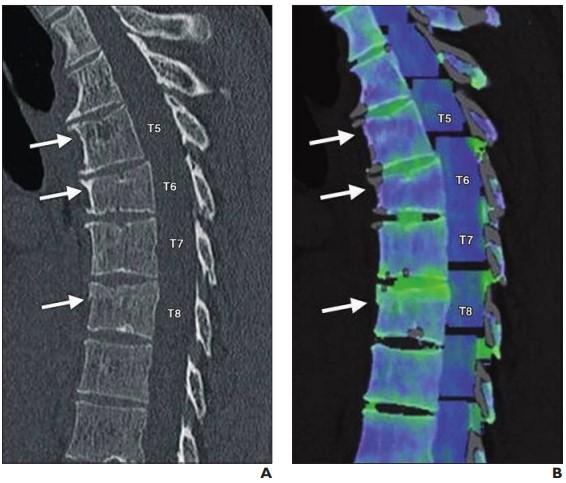
In terms of the five body systems they categorized, the musculoskeletal system accounted for the greatest number of studies wherein DECT potentially altered management (266/298 cases)–the most common use to confirm gout (185/266).
And although DECT was not noted in 2,272 reports (71.9%), compared with conventional CT alone, DECT findings avoided 162-191 recommended follow-up MRI examinations, 21-28 CT examinations, and 2-25 ultrasound examinations.
Meanwhile, DECT findings did prompt one additional recommended interventional angiography procedure, one ventilation-perfusion scan, and one imaging-guided biopsy.
Ultimately, for the Vancouver General ED in the year 2016, “DECT findings led to a decrease in recommended follow-up imaging examinations totaling an estimated $52,991.53-61,598.44,” Wong and colleagues concluded.
Acknowledging a future need to evaluate how referring clinicians adapt to DECT, as well as how much they trust any added value, the authors of this AJR article added that Vancouver General has since completed the implementation of DECT acquisition for all CT examinations performed in the ED.
###
Founded in 1900, the American Roentgen Ray Society (ARRS) is the first and oldest radiological society in North America, dedicated to the advancement of medicine through the profession of radiology and its allied sciences. An international forum for progress in medical imaging since the discovery of the x-ray, ARRS maintains its mission of improving health through a community committed to advancing knowledge and skills with an annual scientific meeting, monthly publication of the peer-reviewed American Journal of Roentgenology (AJR), quarterly issues of InPractice magazine, AJR Live Webinars and Podcasts, topical symposia, print and online educational materials, as well as awarding scholarships via The Roentgen Fund®.


