Super-Resolution RNA Imaging in Live Cells
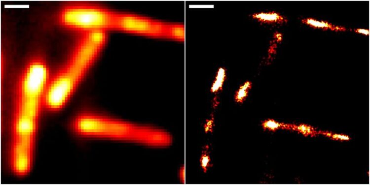
Abbildungen von Darmbakterien mit konventioneller Epifluoreszenzmikroskopie (links) und höchstauflösender Einzelmolekül-Lokalisationsmikroskopie unter Einsatz des neuen RhoBAST-Farbstoff-Marker-Komplexes zur Fluoreszenzmarkierung. Skalenbalken: 1 µm.
Universität Heidelberg/Karlsruher Institut für Technologie
Innovative method provides new insight into molecular processes involving RNA.
Ribonucleic acid (RNA) is key to various fundamental biological processes. It transfers genetic information, translates it into proteins or supports gene regulation. To achieve a more detailed understanding of the precise functions it performs, researchers based at Heidelberg University and at the Karlsruhe Institute of Technology (KIT) have devised a new fluorescence imaging method which enables live-cell RNA imaging with unprecedented resolution.
The method is based on a novel molecular marker called Rhodamine-Binding Aptamer for Super-Resolution Imaging Techniques (RhoBAST). This RNA-based fluorescence marker is used in combination with the dye rhodamine. Due to their distinctive properties, marker and dye interact in a very specific way, which makes individual RNA molecules glow. They can then be made visible using single-molecule localisation microscopy (SMLM), a super-resolution imaging technique. Due to a lack of suitable fluorescence markers, direct observation of RNA via optical fluorescence microscopy has been severely limited to date.
RhoBAST was developed by researchers from the Institute of Pharmacy and Molecular Biotechnology (IPMB) at Heidelberg University and the Institute of Applied Physics (APH) at KIT. The marker created by them is genetically encodable, which means that it can be fused to the gene of any RNA produced by a cell. RhoBAST itself is non-fluorescent, but lights up a cell-permeable rhodamine dye by binding to it in a very specific way. “This leads to a dramatic increase in fluorescence achieved by the RhoBAST-dye complex, which is a key requirement for obtaining excellent fluorescence images,” explains Dr Murat Sünbül from the IPMB, adding: “However, for super-resolution RNA imaging the marker needs additional properties.”
The researchers discovered that each rhodamine dye molecule remains bound to RhoBAST for approximately one second only before becoming detached again. Within seconds, this procedure repeats itself with a new dye molecule. “It is quite rare to find strong interactions – as between RhoBAST and rhodamine – combined with exceptionally fast exchange kinetics,” says Prof. Dr Gerd Ulrich Nienhaus from the APH. Since rhodamine only lights up after binding to RhoBAST, the constant string of newly emerging interactions between marker and dye results in incessant “blinking”. “This ‘on-off switching’ is exactly what we need for SMLM imaging,” continues Prof. Nienhaus.
At the same time, the RhoBAST system solves yet another important problem. Fluorescence images are collected under laser light irradiation, which destroys the dye molecules over time. The fast dye exchange ensures that photobleached dyes are replaced by fresh ones. This means that individual RNA molecules can be observed for longer periods of time, which can greatly improve image resolution, as Prof. Dr Andres Jäschke, a scientist at the IPMB, explains.
The researchers from Heidelberg and Karlsruhe were able to demonstrate the superb properties of RhoBAST as an RNA marker by visualising RNA structures inside gut bacteria (Escherichia coli) and cultured human cells with excellent localisation precision. “We can reveal details of previously invisible subcellular structures and molecular interactions involving RNA using super-resolution fluorescence microscopy. This will enable a fundamentally new understanding of biological processes,” says Prof. Jäschke.
The research carried out by Murat Sünbül and Andres Jäschke in the context of the study was supported by the German Research Foundation (DFG) and the work performed by Gerd Ulrich Nienhaus was supported by the DFG and the Helmholtz Association. The results were published in the journal “Nature Biotechnology”.
Contact:
Communications and Marketing
Press Office
Phone +49 6221 54-2311
presse@rektorat.uni-heidelberg.de
Wissenschaftliche Ansprechpartner:
Contacts at Heidelberg University:
Dr Murat Sünbül
Prof. Dr Andres Jäschke
Institute of Pharmacy and Molecular Biotechnology
Phone +49 6221 54 4853
msunbul@uni-heidelberg.de
jaeschke@uni-hd.de
Contact at Karlsruhe Institute of Technology:
Prof. Dr Gerd Ulrich Nienhaus
Institute of Applied Physics
Phone +49 721 608 43401
uli.nienhaus@kit.edu
Originalpublikation:
M. Sunbul, J. Lackner, A. Martin, D. Englert, B. Hacene, K. Nienhaus, G. U. Nienhaus, A. Jäschke: Super-resolution RNA imaging using a rhodamine-binding aptamer with fast exchange kinetics, Nature Biotechnology, 11 February 2021 (date of online publication), https://doi.org/10.1038/s41587-020-00794-3
Media Contact
All latest news from the category: Life Sciences and Chemistry
Articles and reports from the Life Sciences and chemistry area deal with applied and basic research into modern biology, chemistry and human medicine.
Valuable information can be found on a range of life sciences fields including bacteriology, biochemistry, bionics, bioinformatics, biophysics, biotechnology, genetics, geobotany, human biology, marine biology, microbiology, molecular biology, cellular biology, zoology, bioinorganic chemistry, microchemistry and environmental chemistry.


Newest articles

A universal framework for spatial biology
SpatialData is a freely accessible tool to unify and integrate data from different omics technologies accounting for spatial information, which can provide holistic insights into health and disease. Biological processes…

How complex biological processes arise
A $20 million grant from the U.S. National Science Foundation (NSF) will support the establishment and operation of the National Synthesis Center for Emergence in the Molecular and Cellular Sciences (NCEMS) at…

Airborne single-photon lidar system achieves high-resolution 3D imaging
Compact, low-power system opens doors for photon-efficient drone and satellite-based environmental monitoring and mapping. Researchers have developed a compact and lightweight single-photon airborne lidar system that can acquire high-resolution 3D…

