Simplified diagnosis of rare eye diseases
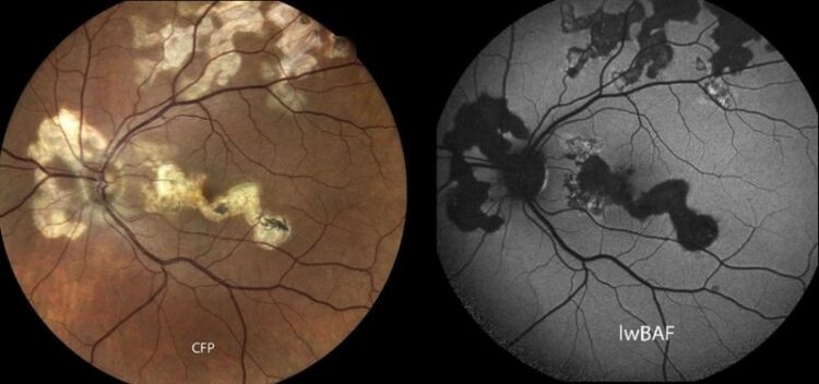
Rare serpiginous chorioretinopathy: The ocular fundus photograph (left) shows areas of scarring in a light yellow tone. Active inflammatory lesions usually appear as light-colored areas on fundus autofluorescence (right).
(c) University Hospital Bonn (UKB)
Uveitis experts provide an overview of an underestimated imaging technique.
Uveitis is a rare inflammatory eye disease. Posterior and panuveitis in particular are associated with a poor prognosis and a protracted course of the disease. Diagnosis and monitoring can be challenging for healthcare professionals. Fundus autofluorescence (FAF) is a fast and non-invasive imaging technique that supports this. Researchers from the University Hospital Bonn and the University of Bonn, together with experts from Berlin, Münster and Mannheim, have drafted a review on how FAF can facilitate the diagnosis and monitoring of posterior uveitis and panuveitis. The results have now been published in the journal “Biomolecules”.
Uveitis is a rare inflammatory disease of the choroid of the eye, which lies between the retina and the sclera. “Depending on the inflamed anatomical structure, this disease can be divided into the subtypes anterior, intermediate, posterior and panuveitis. The exact diagnosis of posterior uveitis and panuveitis can be challenging, as there are many different and sometimes extremely rare subtypes,” explains Dr. Maximilian Wintergerst from the Eye Clinic at the University Hospital Bonn (UKB), who also conducts research at the University of Bonn. In the review, the researchers from Bonn, Berlin, Münster, and Mannheim now show how imaging using fundus autofluorescence (FAF) supports the diagnosis and monitoring of some posterior uveitis forms.
FAF provides indications of active inflammation
Fundus autofluorescence is a non-invasive method for imaging the fundus of the eye. “Using light of a precisely defined wavelength, so-called fluorophores in the tissue of the eye are stimulated to glow. The distribution of these fluorophores, the intensity of the light signal, and certain resulting light patterns can provide information about the underlying form of uveitis,” explains Wintergerst. In unclear cases, this can help to make the correct diagnosis. “In addition, the autofluorescence signal can also provide us with information on the current state of inflammation in certain forms of uveitis. For example, brightly illuminated areas in the retina are sometimes associated with active inflammation, while darker areas can indicate inactive inflammation,” adds Dr. Matthias Mauschitz, Head of the Uveitis Clinic at the UKB.
The wavelength used influences the result
“Depending on the wavelength used, the autofluorescence signal from the retina and choroid can differ significantly. Depending on the excitation wavelength, lesions can be imaged at different depths and therefore in different areas,” explains Mauschitz. In addition to their review, the researchers included a case series in which they compared the autofluorescence of different wavelengths. Overall, they found that the combination of different wavelengths can provide additional information about the underlying form of uveitis.
Combination of different wavelengths provides additional information
With their work, the research team would like to draw attention to autofluorescence imaging, which is very helpful in some forms of uveitis, and highlight new approaches for future research, such as the combination of autofluorescence imaging of different wavelengths. “Fundus autofluorescence plays an important role in the diagnosis and monitoring of posterior uveitis and panuveitis. In some specific subtypes of uveitis, it can also provide important indications of a flare-up of inflammatory activity,” summarizes Wintergerst.
Involved institutions and financing:
In addition to the UKB and the University of Bonn, the Sankara Eye Hospital Shimoga in India, the Grischun Eye Center in Switzerland, the St. Franziskus-Hospital Münster, the University of Duisburg-Essen, the Charité in Berlin and the University Medical Center Mannheim are also involved. The project was supported by funds from the BONFOR GEROK program of the medical faculty of the University of Bonn (grant number O-137.0028) and the Ernst and Berta Grimmke Foundation (grant number 3/22).
Publication:
Matthias M. Mauschitz, Markus Zeller et al: Fundus Autofluorescence in Posterior and Panuveitis – An Under-Estimated Imaging Technique: A Review and Case Series; Biomolecules; DOI: 10.3390/biom14050515
Link: https://www.mdpi.com/2218-273X/14/5/515
Press contact:
Dr. Inka Väth
Deputy Press Officer at the University Hospital Bonn (UKB)
Communications and Media Office at Bonn University Hospital
Phone: (+49) 228 287-10596
E-mail: inka.vaeth@ukbonn.de
About Bonn University Hospital: The UKB treats around 500,000 patients per year, employs around 9,000 staff and has total assets of 1.6 billion euros. In addition to the 3,500 medical and dental students, 550 people are trained in numerous healthcare professions each year. The UKB is ranked first among university hospitals (UK) in NRW in the science ranking and in the Focus clinic list and has the third highest case mix index (case severity) in Germany. In 2022 and 2023, the F.A.Z. Institute recognized the UKB as the most desirable employer and training champion among public hospitals in Germany.
Wissenschaftliche Ansprechpartner:
Priv.-Doz. Dr. med. Maximilian W. M. Wintergerst
Eye Clinic of the University Hospital Bonn
E-Mail: Maximilian.Wintergerst@ukbonn.de
Originalpublikation:
Matthias M. Mauschitz, Markus Zeller et al: Fundus Autofluorescence in Posterior and Panuveitis – An Under-Estimated Imaging Technique: A Review and Case Series; Biomolecules; DOI: 10.3390/biom14050515
Weitere Informationen:
https://www.mdpi.com/2218-273X/14/5/515 Publikation
Media Contact
All latest news from the category: Health and Medicine
This subject area encompasses research and studies in the field of human medicine.
Among the wide-ranging list of topics covered here are anesthesiology, anatomy, surgery, human genetics, hygiene and environmental medicine, internal medicine, neurology, pharmacology, physiology, urology and dental medicine.


Newest articles

Faster, more energy-efficient way to manufacture an industrially important chemical
Zirconium combined with silicon nitride enhances the conversion of propane — present in natural gas — needed to create in-demand plastic, polypropylene. Polypropylene is a common type of plastic found…

Energy planning in Ghana as a role model for the world
Improving the resilience of energy systems in the Global South. What criteria should we use to better plan for resilient energy systems? How do socio-economic, technical and climate change related…

Artificial blood vessels could improve heart bypass outcomes
Artificial blood vessels could improve heart bypass outcomes. 3D-printed blood vessels, which closely mimic the properties of human veins, could transform the treatment of cardiovascular diseases. Strong, flexible, gel-like tubes…

