Visualizing cell structures in three dimensions in mere minutes
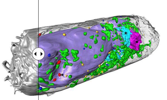
Human lung epithelium cell 24 hours after SARS-CoV-2 viral infection. Hijacked cellular organelles are labelled with an asterisk.
Credit: Venera Weinhardt (COS)
Heidelberg researchers are working on a rapid process for 3D imaging of cells.
Viral pathogens like the SARS-CoV-2 coronavirus change the interior structure of the cells they infect. These changes occur at the level of individual cell components – the organelles – and can provide information on how viral diseases develop. Extremely powerful imaging techniques are needed to visualise them, but such methods are very data- and time-intensive. A German-American research team under the direction of Dr Venera Weinhardt at the Centre for Organismal Studies (COS) of Heidelberg University recently optimised a special X-ray process – known as soft X-ray tomography – to deliver high-resolution three-dimensional images of entire cells and their molecular structure in just a few minutes.
“Scanning electron microscopes are preferred in cell imaging because they provide extremely sharp nanoscale images,” explains Venera Weinhardt, a post-doc at the COS and the Lawrence Berkeley National Laboratory in Berkeley (USA). “But this technology takes a good week to scan an individual cell. It also generates an enormous amount of data that is daunting to analyse and interpret. Using soft X-ray tomography, we get usable results within five to ten minutes.” High throughput is extremely important for studying numerous cells, according to molecular virologist Prof. Dr Ralf Bartenschlager, whose department at Heidelberg University Hospital is collaborating with Dr Weinhardt on imaging cellular changes associated with viral infections. In tissue, the scientist adds, often only some of the cells are infected. Only those cells provide information on the changes that result directly from the infection. Looking for these cells with a scanning electron microscope, however, is not possible.
The procedure known as soft X-ray tomography (SXT) has already been used to successfully detect single virus particles – called virions – of different types of viruses and their associated changes in cells. Now the researchers used the technology to study cell cultures infected with SARS-CoV-2 from lung and kidney tissue. Soft X-rays allowed them to image complete cells and their structure in three dimensions in five to ten minutes. The researchers were further able to detect clusters of SARS-CoV-2 particles on cell surfaces as well as identify virus-associated changes in the cell’s interior. Structures were revealed that possibly enable the replication and spread of the virus.
According to Dr Weinhardt, the team’s success largely hinged on the technology allowing them to study fixed cells, i.e. cells that had been chemically treated to deactivate the virus. Typically, in soft X-ray tomography, like in electron tomography, flat lattice structures are used as holders. When they are tilted, the thickness of the samples can change, making some cell structures appear blurry. “Blind” spots also occur because the flat shape of the holder prevents the cells from being scanned at all angles. Another dilemma is that the samples can adhere to the lattice or spread out, requiring multiple tomograms to visualise the entire cell. “To get around this problem, we switched over to cylindrical thin-wall glass capillaries to hold the samples. During microscopy, the samples can be rotated a full 360 degrees and scanned from all angles,” explains the researcher. The team is now working on further refining sample preparation techniques, automating the analysis of the 3D image data, and developing a laboratory version of a soft X-ray microscope.
Funding for the research in Heidelberg was provided by the German Research Foundation (DFG), the European Research Council (ERC) and the “Horizon 2020” Framework Programme of the European Union. The results of the research were published in the journal “Cell Reports Methods”.
Journal: Cell Reports Methods
DOI: 10.1016/j.crmeth.2021.100117
Article Title: Using soft X-ray tomography for rapid whole-cell quantitative imaging of SARS-CoV-2-infected cells
Article Publication Date: 22-Nov-2021
Media Contact
Ute Mueller-Detert
Heidelberg University
ute.mueller-detert@rektorat.uni-heidelberg.de
Office: 004-962-2154 x19017
Expert Contact
Dr Venera Weinhardt
Centre for Organismal Studies
venera.weinhardt@cos.uni-heidelberg.de
Media Contact
All latest news from the category: Life Sciences and Chemistry
Articles and reports from the Life Sciences and chemistry area deal with applied and basic research into modern biology, chemistry and human medicine.
Valuable information can be found on a range of life sciences fields including bacteriology, biochemistry, bionics, bioinformatics, biophysics, biotechnology, genetics, geobotany, human biology, marine biology, microbiology, molecular biology, cellular biology, zoology, bioinorganic chemistry, microchemistry and environmental chemistry.


Newest articles

High-energy-density aqueous battery based on halogen multi-electron transfer
Traditional non-aqueous lithium-ion batteries have a high energy density, but their safety is compromised due to the flammable organic electrolytes they utilize. Aqueous batteries use water as the solvent for…

First-ever combined heart pump and pig kidney transplant
…gives new hope to patient with terminal illness. Surgeons at NYU Langone Health performed the first-ever combined mechanical heart pump and gene-edited pig kidney transplant surgery in a 54-year-old woman…

Biophysics: Testing how well biomarkers work
LMU researchers have developed a method to determine how reliably target proteins can be labeled using super-resolution fluorescence microscopy. Modern microscopy techniques make it possible to examine the inner workings…

