Formation of cellulose fibers tracked for the first time
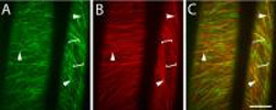
Molecules of cellulose synthase, the enzyme that produces cellulose, follow microtubule “tracks” in growing plant cells. Each image in this set consists of 30 frames, taken at 10 second intervals ...
However, recent work from the Carnegie Institution’s Department of Plant Biology and Stanford University describes the first real-time observations of cellulose fiber formation. The research, published in the April 20 online issue of Science Express, provides the first clear evidence for a functional connection between synthesis of the cell wall and an array of protein fibers–called microtubules–that help to shape growing plant cells from the inside.
“The more we understand about cellulose, the easier it will be to modify it,” said Chris Somerville, director of the Carnegie department. “With this knowledge, we are one step closer to designing energy-rich biofuel crops and improved fiber crops.”
Cellulose fibers make up a significant portion of the dry weight of most plants. Because the fibers can be broken down into the sugar glucose, which can then be converted into ethanol and other biofuels, there are huge incentives to learn more about how plants produce and modify the molecule. Cellulose is also the main constituent of cotton, paper, wood, and animal feeds such as hay.
Somerville, along with colleague David Ehrhardt and Stanford graduate student Alex Paredez, engineered plants to produce a fluorescent version of cellulose synthase, the enzyme that creates cellulose fibers. They also included a fluorescent version of tubulin, the protein from which microtubules are built. Using an imaging technique that can track the motion of single fluorescent molecules, the researchers found that cellulose synthase moves along “tracks” defined by the microtubules. When the microtubule tracks were disrupted with specific drugs, the cellulose synthase molecules kept moving, but they followed different, less directed patterns.
“Many scientists had suspected a relationship between cellulose synthase and microtubules, but the exact nature of the interaction was hard to pinpoint,” Ehrhardt said. “The microtubules act as more than a general scaffold for organizing the cell wall; individual elements of the microtubule array appear to actively direct the pattern of the cellulose fibers. This work should set a long-standing discussion to rest.”
Media Contact
More Information:
http://www.stanford.eduAll latest news from the category: Life Sciences and Chemistry
Articles and reports from the Life Sciences and chemistry area deal with applied and basic research into modern biology, chemistry and human medicine.
Valuable information can be found on a range of life sciences fields including bacteriology, biochemistry, bionics, bioinformatics, biophysics, biotechnology, genetics, geobotany, human biology, marine biology, microbiology, molecular biology, cellular biology, zoology, bioinorganic chemistry, microchemistry and environmental chemistry.


Newest articles

A universal framework for spatial biology
SpatialData is a freely accessible tool to unify and integrate data from different omics technologies accounting for spatial information, which can provide holistic insights into health and disease. Biological processes…

How complex biological processes arise
A $20 million grant from the U.S. National Science Foundation (NSF) will support the establishment and operation of the National Synthesis Center for Emergence in the Molecular and Cellular Sciences (NCEMS) at…

Airborne single-photon lidar system achieves high-resolution 3D imaging
Compact, low-power system opens doors for photon-efficient drone and satellite-based environmental monitoring and mapping. Researchers have developed a compact and lightweight single-photon airborne lidar system that can acquire high-resolution 3D…

