In one sweep: Visualization of protein mobility in the synapse
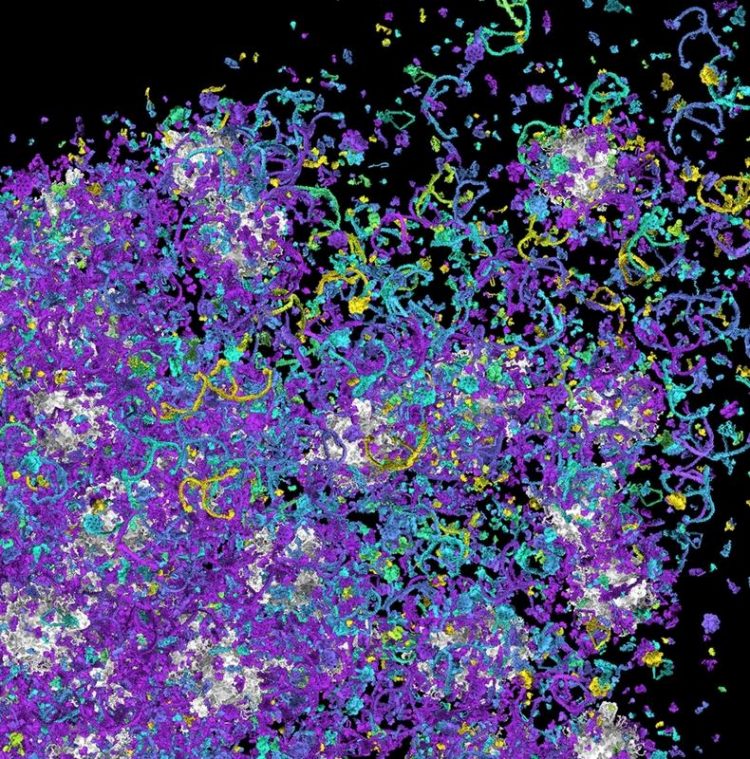
Snapshot of protein mobility in the synapse. Source: Reshetniak and co-authors
All functions of the neural system, including body movements, sensory reactions, and information processing in the brain, depend on the constant and controlled communication between nerve cells.
The contact sites between nerve cells, the synapses, have been extensively studied over the last decades, extending our understanding of brain functioning on a molecular level.
Regarding the identity, numbers, and positions of protein molecules present in the synapses a very detailed view already exists. In contrast, very limited information is available about the dynamics and mobility of these proteins in living synapses.
Knowledge of the mobility rates of the key synaptic proteins would help to understand how these proteins are involved in synaptic signal transduction, and what the possible mechanisms are that regulate their distribution in the synapses.
A team of scientists around Prof. Dr. Silvio O. Rizzoli, Director of the Institute of Neuro- and Sensory Physiology of the University Medical Center Göttingen (UMG), and speaker of the Board of the Center for Biostructural Imaging of Neurodegener-ation (BIN), and Prof. Dr. Sarah Köster, Institute for X-Ray Physics at the Georg-August-University Göttingen, was able to generate the first visualizations of move-ment of 45 proteins in a cell simultaneously, thus demonstrating the realistic mobility of thousands of protein molecules represented in their realistic shapes and sizes within a synapse.
The study uncovers several correlations of mobility parameters to the presence of different building blocks of the proteins, namely to different amino acids in the protein sequence or to the presence of particular nucleotides in the protein-encoding mRNA sequence.
The results of the scientists engaged in the Cluster of Excellence Multiscale Bioimaging (MBExC) and the Göttingen Collaborative Research Centers 1286 and 1190 are summarized in a video animation, and have been published in the renowned scientific journal “The EMBO Journal” on July 6, 2020.
Original publication: A comparative analysis of the mobility of 45 proteins in the synaptic bouton. Sofiia Reshetniak, Jan-Eike Ußling, Eleonora Perego, Burkhard Rammner, Thomas Schikorski, Eugenio F Fornasiero, Sven Truckenbrodt, Sarah Köster, Silvio O Rizzoli, EMBO J, 06.07.2020, doi: 10.15252/embj.2020104596
Link to the video animation:
https://bin.umg.eu/news-details/news-detail/detail/news/mobility-of-proteins/
Animation: A view of soluble protein movement in the synapse. Only synaptic vesicles (grey shapes) and soluble proteins are shown, the plasma membrane surrounding the synapse is transparent. Each colored shape is a single protein molecule, with the molecules of the same protein type having the same color and shape. (Source: Reshetniak and co-authors)
“Our entire data set can be exploited worldwide by laboratories specialized in neuronal and synaptic modeling for generation of multi-reaction models of the synapse”, says Prof. Rizzoli, Director of the Institute of Neuro- and Sensory Physiology and senior author of the study.
“The use of such datasets will reveal important details about synaptic activity in health and neurological or neurodegenerative diseases”, explains Rizzoli. These visualizations can also be appreciated by the general public, and might become a useful educational tool to demonstrate the complexity and dynamics of the cells at the nanoscale.
Research results in Detail
While mobility of a few individual proteins was assessed in the past, it remains challenging to measure mobility of multiple different proteins in a complex environment as a living synapse. The aim of the present study was therefore to obtain a comparative data set on the mobility of up to 50 different proteins in synapses and axons of living neurons in a brain region (hippocampus) involved in memory formation.
“We aimed to understand how protein distribution and mobility in the synapses is regulated, and what the contribution of the synaptic vesicle cluster is, as well as to analyse the connections between protein mobility and various functional protein parameters”, explains Sofiia Reshetniak, PhD student at the Institute of Neuro- and Sensory Physiology and first author of the study.
By combining fluorescence recovery after photobleaching, particle tracking, electron microscopy, and in silico modelling, the mobility of 45 different proteins in living neurons has been analysed and their movement speeds in the form of diffusion coefficients in synapses and axons has been obtained.
“Interestingly, the protein size has only a very limited effect on the mobility of proteins”, says Prof. Köster, one of the senior authors of the study. “Instead, protein association with the synaptic vesicle cluster plays a much more important role”, Prof. Köster adds.
The Göttingen Cluster of Excellence Multiscale Bioimaging: From Molecular Machines to Networks of Excitable Cells (MBExC) is funded since January 2019 in the framework of the Excellence Strategy of the German Federal and State Governments. Applying a unique and multiscale approach, MBExC investigates the disease-relevant functional units of electrically active cells of heart and brain, from the molecular to the organ level.
The MBExC unites numerous partners from the university and extra-university institutions in Göttingen. The overall goal: to understand the relationship between heart and brain diseases, to link basic and clinical research, and thus to develop new therapeutic and diagnostic approaches with social implications.
Picture caption: Snapshot of protein mobility in the synapse. A region within the synapse at the edge of the synaptic vesicle cluster is shown. Organelles in grey represent synaptic vesicles (each ~40 nm in diameter). Various shapes correspond to different protein molecules, with each protein species having its own shape, as extracted from stuctural studies. The proteins are colored based on the speed of movement of every individual molecule in every position, from dark violet (the slowest one) to yellow (the fastest). Most proteins move slowly next to the synaptic vesicles, and are much faster in the vesicle-free region. (Source: Reshetniak and co-authors)
Further Information
about Rizzoli Lab: http://rizzoli-lab.de/
about the Institute for X-Ray Physics: https://www.uni-goettingen.de/en/91107.html
about MBExC: https://mbexc.de/
about CRC1286: https://www.sfb1286.de/
about CRC1190: https://www.sfb1190.de/
Further Information
University Medical Center Göttingen, Georg-August-University
Institute of Neuro- and Sensory Physiology
Prof. Dr. Silvio O. Rizzoli
Humboldtallee 23, D-37073 Göttingen
Phone: +49 (0) 551 / 39-5911, srizzoli@gwdg.de
Georg-August-University Göttingen
Institute for X-Ray Physics, RG Cellular Biophysics
Prof. Dr. Sarah Köster
Friedrich-Hund-Platz 1, D-37077 Göttingen
Phone: +49 (0)551 / 39-29429, sarah.koester@phys.uni-goettingen.de
Original publication: A comparative analysis of the mobility of 45 proteins in the synaptic bouton. Sofiia Reshetniak, Jan-Eike Ußling, Eleonora Perego, Burkhard Rammner, Thomas Schikorski, Eugenio F Fornasiero, Sven Truckenbrodt, Sarah Köster, Silvio O Rizzoli, EMBO J, 06.07.2020, doi: 10.15252/embj.2020104596
Media Contact
All latest news from the category: Life Sciences and Chemistry
Articles and reports from the Life Sciences and chemistry area deal with applied and basic research into modern biology, chemistry and human medicine.
Valuable information can be found on a range of life sciences fields including bacteriology, biochemistry, bionics, bioinformatics, biophysics, biotechnology, genetics, geobotany, human biology, marine biology, microbiology, molecular biology, cellular biology, zoology, bioinorganic chemistry, microchemistry and environmental chemistry.


Newest articles

A universal framework for spatial biology
SpatialData is a freely accessible tool to unify and integrate data from different omics technologies accounting for spatial information, which can provide holistic insights into health and disease. Biological processes…

How complex biological processes arise
A $20 million grant from the U.S. National Science Foundation (NSF) will support the establishment and operation of the National Synthesis Center for Emergence in the Molecular and Cellular Sciences (NCEMS) at…

Airborne single-photon lidar system achieves high-resolution 3D imaging
Compact, low-power system opens doors for photon-efficient drone and satellite-based environmental monitoring and mapping. Researchers have developed a compact and lightweight single-photon airborne lidar system that can acquire high-resolution 3D…

