Software tool finds ’needles’ in data ’haystacks’
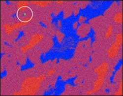
X-ray data collected with a scanning electron microscope from a nickel-aluminum alloy. Pure aluminum is represented by blue, pure nickel by red and nickel-aluminum alloys by colors in between. The green dot in the upper left shows a contaminant particle of chromium identified with the NIST software that occupied only one pixel of the microscope’s scanning area. The sample measures about 160 micrometers across. Image credit: D. Bright, D. Newbury/NIST
When looking for a needle in a haystack, it’s helpful to know what a needle looks like. A new software tool developed by researchers at the National Institute of Standards and Technology (NIST) makes it possible to find chemical ’needles’ in data ’haystacks’ without having to know anything about the ’needle’ in advance.
The NIST software should be especially useful for analyzing ultrapure metals–recently shown to have superior strength, corrosion-resistance and other properties–and for monitoring nanoscale semiconductor fabrication. Commercial X-ray detector manufacturers already have included the method used in the software into their products.
Described in the November issue of the Journal of Microscopy*, the software works with scanning electron microscopes (SEMs) and improves the analysis of X-ray data. SEMs raster a beam of electrons across a sample and then detect X-rays emitted in response. X-rays of specific energies (the equivalent of colors for visible light) are emitted by specific elements, making SEMs an excellent tool for mapping the chemical composition of samples. The lateral and depth resolutions of SEM/X-ray analysis range from 100 nanometers to 5 micrometers, depending on specimen composition and SEM beam energy.
Newer detectors—some developed with NIST funding—respond so fast that data across the entire spectrum of X-ray energies can be recorded for every pixel scanned. Typically, these data are analyzed to show only the sample’s major constituents. The NIST software analyzes the data a step further by identifying the X-ray energy with the highest intensity for each pixel rather than for the sample as a whole. Using the software with a nickel-aluminum sample, the NIST researchers identified chromium and copper contaminant particles that occupied just a single pixel and were not “visible” with the SEM’s usual data interpretation tools.
Media Contact
More Information:
http://www.nist.govAll latest news from the category: Information Technology
Here you can find a summary of innovations in the fields of information and data processing and up-to-date developments on IT equipment and hardware.
This area covers topics such as IT services, IT architectures, IT management and telecommunications.


Newest articles

High-energy-density aqueous battery based on halogen multi-electron transfer
Traditional non-aqueous lithium-ion batteries have a high energy density, but their safety is compromised due to the flammable organic electrolytes they utilize. Aqueous batteries use water as the solvent for…

First-ever combined heart pump and pig kidney transplant
…gives new hope to patient with terminal illness. Surgeons at NYU Langone Health performed the first-ever combined mechanical heart pump and gene-edited pig kidney transplant surgery in a 54-year-old woman…

Biophysics: Testing how well biomarkers work
LMU researchers have developed a method to determine how reliably target proteins can be labeled using super-resolution fluorescence microscopy. Modern microscopy techniques make it possible to examine the inner workings…

