3D images of cancer cells in the body: Medical physicists from Halle present new method
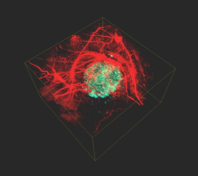
A picture of a tumor (green) generated with the newly developed technique. Jan Laufer
Clinicians and scientists are in need of a better understanding of cancer cells and their properties in order to provide targeted cancer treatment. Individual cancer cells are often examined in test tubes before the findings are tested in living organisms. “
Our aim is to visualise cancer cells inside the living body to find out how they function, how they spread and how they react to new therapies,” says medical physicist Professor Jan Laufer from MLU. He specialises in the field of photoacoustic imaging, a process that uses ultrasound waves generated by laser beams to produce high-resolution, three-dimensional images of the body’s interior.
“The problem is that tumour cells are transparent. This makes it difficult to use optical methods to examine tumours in the body,” explains Laufer whose research group has developed a new method to solve this problem: First the scientists introduce a specific gene into the genome of the cancer cells.
“Once inside the cells, the gene produces a phytochrome protein, which originates from plants and bacteria. There it serves as a light sensor,” Laufer continues. In the next step, the researchers illuminate the tissue with short pulses of light at two different wavelengths using a laser. Inside the body, the light pulses are absorbed and converted into ultrasonic waves.
These waves can then be measured outside the organism and two images of the body's interior can be reconstructed based on this data. “The special feature of phytochrome proteins is that they alter their structure and thus also their absorption properties depending on the wavelength of the laser beams.
This results in changes to the amplitude of the ultrasound waves that are generated in the tumour cells. None of the other tissue components, for example blood vessels, have this property – their signal remains constant,” Laufer says. By calculating the difference between the two images, a high-resolution, three-dimensional image of the tumour cells is created, which is free of the otherwise overwhelming background contrast.
The development of Halle's medical physicists can be applied to a wide range of applications in the preclinical research and the life sciences. In addition to cancer research, the method can be used to observe cellular and genetic processes in living organisms.
About the study:
J. Märk et. al, Dual-wavelength 3D photoacoustic imaging of mammalian cells using a photoswitchable phytochrome reporter protein. Communication Physics 2018, 1, 3. DOI: 10.1038/s42005-017-0003-2
Media Contact
More Information:
http://www.uni-halle.deAll latest news from the category: Health and Medicine
This subject area encompasses research and studies in the field of human medicine.
Among the wide-ranging list of topics covered here are anesthesiology, anatomy, surgery, human genetics, hygiene and environmental medicine, internal medicine, neurology, pharmacology, physiology, urology and dental medicine.


Newest articles

A universal framework for spatial biology
SpatialData is a freely accessible tool to unify and integrate data from different omics technologies accounting for spatial information, which can provide holistic insights into health and disease. Biological processes…

How complex biological processes arise
A $20 million grant from the U.S. National Science Foundation (NSF) will support the establishment and operation of the National Synthesis Center for Emergence in the Molecular and Cellular Sciences (NCEMS) at…

Airborne single-photon lidar system achieves high-resolution 3D imaging
Compact, low-power system opens doors for photon-efficient drone and satellite-based environmental monitoring and mapping. Researchers have developed a compact and lightweight single-photon airborne lidar system that can acquire high-resolution 3D…

