New microscopy concept enters into force
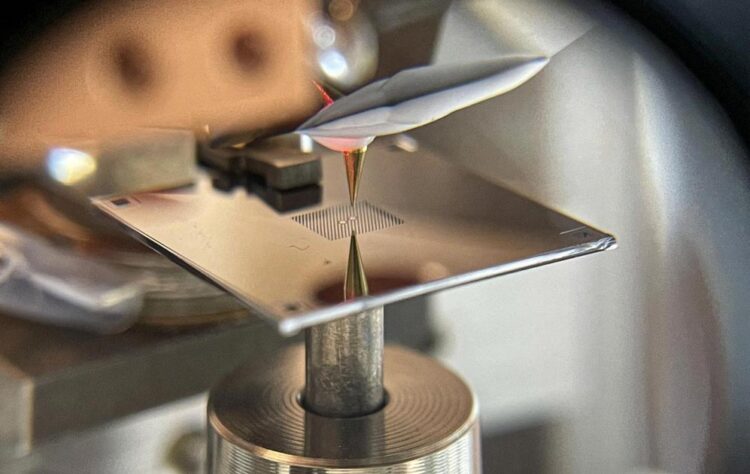
The separation of the islands is around half a millimetre.
Credit: David Hälg and Shobhna Misra, ETH Zurich
The first demonstration of an approach that inverts the standard paradigm of scanning probe microscopy raises the prospect of force sensing at the fundamental limit.
The development of scanning probe microscopes in the early 1980s brought a breakthrough in imaging, throwing open a window into the world at the nanoscale. The key idea is to scan an extremely sharp tip over a substrate and to record at each location the strength of the interaction between tip and surface. In scanning force microscopy, this interaction is — as the name implies — the force between tip and structures on the surface.
This force is typically determined by measuring how the dynamics of a vibrating tip changes as it scans over objects deposited on a substrate. A common analogy is tapping a finger across a table and sensing objects placed on the surface. A team led by Alexander Eichler, Senior Scientist in the group of Prof. Christian Degen at the Departement of Physics of ETH Zurich, turned this paradigm upside down. Writing in Physical Review Applied, they report the first scanning force microscope in which the tip is at rest while the substrate with the samples on it vibrates.
Tail wagging the dog
Doing force microscopy by ‘vibrating the table under the finger’ may look like making the entire procedure a whole lot more complicated. In a sense it does. But mastering the complexity of this inverted approach comes with great payoff. The new method promises to push the sensitivity of force microscopy to its fundamental limit, beyond what can be expected from further improvements of the conventional ‘finger tapping’ approach.
The key to the superior sensitivity is the choice of substrate. The ‘table’ in the experiments of Eichler, Degen and their co- workers is a perforated membrane made of silicon nitride, a mere 41 nm in thickness. Collaborators of the ETH physicists, the group of Albert Schliesser at the University of Copenhagen in Denmark, have established these low- mass membranes as outstanding nanomechanical resonators with extreme ‘quality factors’. That is, that once the membrane is tipped on, it vibrates millions of times, or more, before coming to rest. Given these exquisite mechanical properties, it becomes advantageous to vibrate the ‘table’ rather than the ‘finger’. At least in principle.
New concept put to practice
Translating this theoretical promise into experimental capability is the objective of an ongoing project between the groups of Degen and Schliesser, with theory support from Dr. Ramasubramanian Chitra and Prof. Oded Zilberberg of the Institute for Theoretical Physics at ETH Zurich. As a milestone on that journey, the experimental teams have now demonstrated that the concept of membrane- based scanning force microscopy works in a real device.
In particular, they showed that neither loading the membrane with samples nor bringing the tip to within a distance of a few nanometres compromises the exceptional mechanical properties of the membrane. However, once the tip approaches the sample even closer, the frequency or amplitude of the membrane changes. To be able to measure these changes, the membrane features not only an island where tip and sample interact, but also a second one — mechanically coupled to the first — from where a laser beam can be partially reflected, to provide a sensitive optical interferometer.
Quantum is the limit
Putting this setup to work, the team successfully resolved gold nanoparticles and tobacco mosaic viruses. These images serve as a proof of principle for the novel microscopy concept, but they do not yet push the capabilities into new territory. But the destination is just there. The researchers plan to combine their novel approach with a technique known as magnetic resonance force microscopy (MRFM), to enable magnetic resonance imaging (MRI) with a resolution of single atoms, thus providing unique insight, for example, into viruses.
Atomic-scale MRI would be another breakthrough in imaging, combining ultimate spatial resolution with highly specific physical and chemical information about the atoms imaged. For the realization of that vision, a sensitivity close to the fundamental limit given by quantum mechanics is needed. The team is confident that they can realise such a ‘quantum-limited’ force sensor, through further advances in both membrane engineering and measurement methodology. With the demonstration that membrane-based scanning force microscopy is possible, the ambitious goal has now come one big step closer.
Media Contact
All latest news from the category: Physics and Astronomy
This area deals with the fundamental laws and building blocks of nature and how they interact, the properties and the behavior of matter, and research into space and time and their structures.
innovations-report provides in-depth reports and articles on subjects such as astrophysics, laser technologies, nuclear, quantum, particle and solid-state physics, nanotechnologies, planetary research and findings (Mars, Venus) and developments related to the Hubble Telescope.


Newest articles

Simplified diagnosis of rare eye diseases
Uveitis experts provide an overview of an underestimated imaging technique. Uveitis is a rare inflammatory eye disease. Posterior and panuveitis in particular are associated with a poor prognosis and a…

Targeted use of enfortumab vedotin for the treatment of advanced urothelial carcinoma
New study identifies NECTIN4 amplification as a promising biomarker – Under the leadership of PD Dr. Niklas Klümper, Assistant Physician at the Department of Urology at the University Hospital Bonn…
A novel universal light-based technique
…to control valley polarization in bulk materials. An international team of researchers reports in Nature a new method that achieves valley polarization in centrosymmetric bulk materials in a non-material-specific way…

