New method measures the 3D position of individual atoms
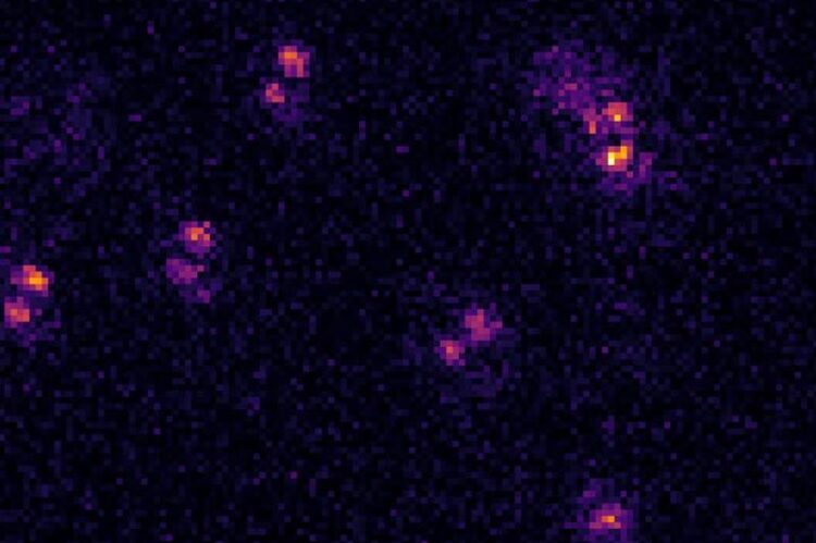
This is how it looks in practice: The different rotational directions of the various “dumbbells” indicate that the atoms lie in different planes.
Abbildung: IAP/Uni Bonn
Since more than a decade it has been possible for physicists to accurately measure the location of individual atoms to a precision of smaller than one thousandth of a millimeter using a special type of microscope. However, this method has so far only provided the x and y coordinates. Information on the vertical position of the atom is lacking. A new method has now been developed that can determine all three spatial coordinates of an atom with one single image. This method – developed by the University of Bonn and University of Bristol – is based on an ingenious physical principle. The study was recently published in the specialist journal Physical Review A.
Anyone who has used a microscope in a biology class to study a plant cell will probably be able to recall a similar situation. It is easy to tell that a certain chloroplast is located above and to the right of the nucleus. But are both of them located on the same plane? Once you adjust the focus on the microscope, however, you see that the image of the nucleus becomes sharper while the image of the chloroplast blurs. One of them must be a little higher and one a little lower than the other. However, this method cannot give us precise details about their vertical positions.
The principle is very similar if you want to observe individual atoms instead of cells. So-called quantum gas microscopy can be used for this purpose. It allows you to straightforwardly determine the x and y coordinates of an atom. However, it is much more difficult to measure its z coordinate, i.e., the distance to the objective lens: In order to find out on what plane the atom is located, multiple images must be taken in which the focus is shifted across various different planes. This is a complex and time-consuming process.
Turning round specks into dumbbells
“We have now developed a method in which this process can be completed in one step,” explains Tangi Legrand from the Institute of Applied Physics (IAP) at the University of Bonn. “To achieve this, we use an effect that has already been known in theory since the 1990s but which had not yet been used in a quantum gas microscope.”
To experiment on the atoms, it is first necessary to cool them down significantly so that they are barely moving. Afterwards, it is possible, for example, to trap them in a standing wave of laser light. They then slip into the troughs of the wave similar to how eggs sit in an egg box. Once trapped, to reveal their position, they are exposed to an additional laser beam, which stimulates them to emit light. The resulting fluorescence shows up in the quantum gas microscope as a slightly blurred, round speck.
“We have now developed a special method to deform the wavefront of the light being emitted by the atom,” explains Dr. Andrea Alberti. The researcher, who has now moved from the IAP to the Max Planck Institute of Quantum Optics in Garching, also participated in the study. “Instead of the typical round specks, the deformed wavefront produces a dumbbell shape on the camera that rotates around itself. The direction in which this dumbbell points is dependent on the distance that the light had to travel from the atom to the camera.”
“The dumbbell thus acts a bit like the needle on a compass, allowing us to read off the z coordinate according to its orientation,” says Prof. Dr. Dieter Meschede. The IAP researcher, whose research group carried out the study, is also a member of the transdisciplinary research area “Matter” at the University of Bonn.
Important for quantum mechanics experiments
The new method makes it possible to precisely determine the position of an atom in three dimensions with one single image. This is important, for example, if you want to carry out quantum mechanics experiments with atoms because it is often essential to be able to precisely control or track their position. This allows researchers to make the atoms interact with one another in the desired way.
Furthermore, the method could also be used to help develop new quantum materials with special characteristics. “For example, we could investigate which quantum mechanical effects occur when atoms are arranged in a certain order,” explains Dr. Carrie Weidner from the University of Bristol. “This would allow us to simulate the properties of three-dimensional materials to some extent without having to synthesize them.”
Participating institutes and funding:
The University of Bonn and the University of Bristol both participated in the study. The research was financed by the German Research Foundation (DFG).
Wissenschaftliche Ansprechpartner:
Tangi Legrand
Nonlinear Quantum Optics Group at the University of Bonn
Tel. +49 228 73-9320
E-mail: legrand@uni-bonn.de
Originalpublikation:
Tangi Legrand, Falk-Richard Winkelmann, Wolfgang Alt, Dieter Meschede, Andrea Alberti and Carrie A. Weidner: Three-dimensional imaging of single atoms in an optical lattice via helical point-spread-function engineering. Phys Rev A, DOI: 10.1103/PhysRevA.109.033304; URL: https://link.aps.org/doi/10.1103/PhysRevA.109.033304
Media Contact
All latest news from the category: Physics and Astronomy
This area deals with the fundamental laws and building blocks of nature and how they interact, the properties and the behavior of matter, and research into space and time and their structures.
innovations-report provides in-depth reports and articles on subjects such as astrophysics, laser technologies, nuclear, quantum, particle and solid-state physics, nanotechnologies, planetary research and findings (Mars, Venus) and developments related to the Hubble Telescope.


Newest articles

A universal framework for spatial biology
SpatialData is a freely accessible tool to unify and integrate data from different omics technologies accounting for spatial information, which can provide holistic insights into health and disease. Biological processes…

How complex biological processes arise
A $20 million grant from the U.S. National Science Foundation (NSF) will support the establishment and operation of the National Synthesis Center for Emergence in the Molecular and Cellular Sciences (NCEMS) at…

Airborne single-photon lidar system achieves high-resolution 3D imaging
Compact, low-power system opens doors for photon-efficient drone and satellite-based environmental monitoring and mapping. Researchers have developed a compact and lightweight single-photon airborne lidar system that can acquire high-resolution 3D…

