SPECT/CT combined with fluorescence imaging detects micrometastases
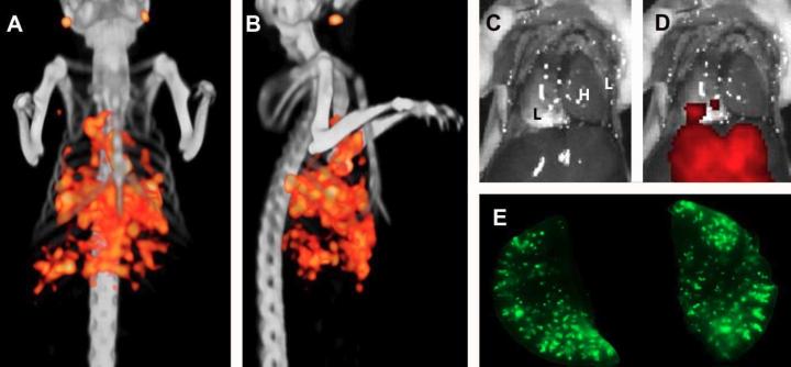
Pulmonary tumor lesions could be visualized with micro-SPECT/CT (coronal view A, sagittal view B). Some physiologic uptake of the tracer in the liver and lymph nodes is observed. After resection of the thoracoabdominal wall, no tumor lesions were visible with the naked eye (C). Fluorescence imaging with the IVIS lumina closed cabinet fluorescence camera system could identify the superficially located tumor lesions (D). Fluorescence imaging with the Odyssey flatbed fluorescence scanner of the resected lungs could visualize numerous pulmonary lesions (E). L = lung, H = heart. Credit: M.C.H. Hekman, Department of Radiology & Nuclear Medicine, Radboudumc, Nijmegen, The Netherlands
Researchers in The Netherlands have demonstrated that combining SPECT/CT and fluorescence imaging could help surgeons differentiate tumor tissue from normal tissue. The research is detailed in the featured basic science article of the May 2017 issue of The Journal of Nuclear Medicine.
The study focused on colorectal cancer (CRC) that metastasized beyond the primary tumor. CRC is the second most common cancer in men and the third most common in women. According to the American Cancer Society, more than 50,000 people in the U.S. are expected to die from the disease in 2017.
“This study shows that micrometastases of colorectal carcinoma can be detected specifically with fluorescence imaging and SPECT/CT before they become visible with the naked eye,” says Marlène C.H. Hekman of the Department of Radiology and Nuclear Medicine at Radboudumc, Nijmegen, The Netherlands.
The researchers used a mouse model to show that pulmonary micrometastases of CRC can be identified with labetuzumab, labeled with both a near-infrared fluorescent dye (IRDye800CW) and a radioactive label (Indium-111). Labetuzumab, developed by Immunomedics, Inc., is an antibody that targets carcinoembryonic antigen (CEA), which is overexpressed in approximately 95 percent of CRC cases. Specifically, it is an investigational anti-CEA-related cell adhesion molecule 5 (CEACAM5) antibody.
Sub-millimeter pulmonary tumor colonies were visualized with both micro-SPECT and fluorescence imaging from the first week of tumor growth. In addition, the study demonstrated that dual-modality imaging can be used to guide resection of tumor lesions.
“Complete tumor resection is crucial for optimal prognosis of cancer patients,” Hekman explains. “Intraoperative imaging can help the surgeon to resect residual disease completely. To be of optimal benefit in the operating room, dual-modality imaging should be able to detect very small tumor lesions that might otherwise be missed.”
She points out, “The study shows that SPECT and fluorescence dual-modality imaging after injection of Indium-111-DTPA-labetuzumab-IRDye800CW is able to detect tiny CEA-expressing nodules. This strategy can also be used for other types of cancer, when a different tumor-targeting antibody is used. It is, therefore, relevant for a broad range of cancer types. Since our findings may be translated to a broad range of tumor types, dual-modality imaging may revolutionize oncological surgery and improve the prognosis of cancer patients.”
###
The authors of Detection of micrometastases using SPECT/fluorescence dual-modality imaging in a CEA-expressing tumor model include Marlene C.H. Hekman, Mark Rijpkema, Desirée Bos, Egbert Oosterwijk, Peter F.A. Mulders, and Otto C. Boerman, Radboudumc, Nijmegen, The Netherlands; and David M. Goldenberg, Immunomedics, Inc., Morris Plains, New Jersey.
Please visit the SNMMI Media Center (http://www.
About the Society of Nuclear Medicine and Molecular Imaging
The Society of Nuclear Medicine and Molecular Imaging (SNMMI) is an international scientific and medical organization dedicated to raising public awareness about nuclear medicine and molecular imaging, a vital element of today's medical practice that adds an additional dimension to diagnosis, changing the way common and devastating diseases are understood and treated and helping provide patients with the best health care possible.
SNMMI's more than 17,000 members set the standard for molecular imaging and nuclear medicine practice by creating guidelines, sharing information through journals and meetings and leading advocacy on key issues that affect molecular imaging and therapy research and practice. For more information, visit http://www.
Media Contact
All latest news from the category: Medical Engineering
The development of medical equipment, products and technical procedures is characterized by high research and development costs in a variety of fields related to the study of human medicine.
innovations-report provides informative and stimulating reports and articles on topics ranging from imaging processes, cell and tissue techniques, optical techniques, implants, orthopedic aids, clinical and medical office equipment, dialysis systems and x-ray/radiation monitoring devices to endoscopy, ultrasound, surgical techniques, and dental materials.


Newest articles

A universal framework for spatial biology
SpatialData is a freely accessible tool to unify and integrate data from different omics technologies accounting for spatial information, which can provide holistic insights into health and disease. Biological processes…

How complex biological processes arise
A $20 million grant from the U.S. National Science Foundation (NSF) will support the establishment and operation of the National Synthesis Center for Emergence in the Molecular and Cellular Sciences (NCEMS) at…

Airborne single-photon lidar system achieves high-resolution 3D imaging
Compact, low-power system opens doors for photon-efficient drone and satellite-based environmental monitoring and mapping. Researchers have developed a compact and lightweight single-photon airborne lidar system that can acquire high-resolution 3D…

