New study decodes one of the living world’s fastest cell movements
Heliozoan Raphidocystis contractilis withdraws its axopodia a few milliseconds after encountering an external stimulus. Researchers from Okayama University, Japan report that microtubule dynamics hold the key to this instant arm shortening
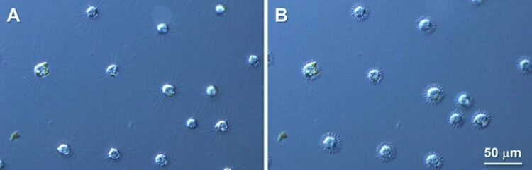
Credit: Motonori Ando from Okayama University
Researchers find the genes and proteins involved in heliozoan arms withdrawal in response to environmental changes, which is one of the fastest examples of cell motility.
Raphidocystis contractilis belongs to Heliozoa, a group of eukaryotes commonly found in fresh, brackish, and sea water. The organisms of this group have finger-like arms—axopodia—which radiate out from their body, giving them a sun-like appearance. Hence, they are also known as “solar worms”. Each axopodium is composed of the proteins, alpha-beta tubulin heterodimers, which form filaments called microtubules. R. contractilis can withdraw its axopodia extremely fast in response to external stimuli. However, the mechanism underlying this rapid arm shortening remains a mystery.
To this end, a team of researchers including Professor Motonori Ando, Dr. Risa Ikeda (both from the Laboratory of Cell Physiology) and Associate Professor Mayuko Hamada (from the Ushimado Marine Institute), of Okayama University, Japan, explored the mechanism involved in one of the fastest cell movements in the living world.
So, where did it all begin? Sharing the motivation behind their study, Professor Ando says, “Recently, a wide variety of heliozoans have been discovered in various hydrospheres in the Okayama Prefecture, making it clear that several species of sun worms inhabit the same environment. We are trying to unravel the mysteries around these protozoans and gradually expand the horizons of our knowledge.”
The authors started their investigation by immunolabelling the tubulin protein and observing its movement before and after axopodial contraction. They found that before shortening, tubulins were arranged systematically all along the length of the axopodia, but after axopodial withdrawal, those swiftly accumulated at the cell surface. This led them to believe that during the rapid axopodial withdrawal, the microtubules broke down into tubulin instantly. However, microtubule degradation is generally not a rapid phenomenon; it progresses rather slowly.
How then, could R. contractilis achieve this change so quickly?
The researchers hypothesized that this was possible if the microtubules split at multiple sites simultaneously. To validate their hypothesis, the authors set out to find the proteins and genes involved in the instant cleavage of microtubules in R. contractilis. Their findings were published online in The Journal of Eukaryotic Microbiology on 21 November 2022.
The researchers performed de novo transcriptome sequencing (analysis of the genes expressed at a particular time in a cell) and identified close to 32,000 genes in R. contractilis. This gene set was most similar to that found in protozoans (which are single-celled organisms), followed by metazoans (multicellular organisms with well-differentiated cells; this includes humans, and other animals).
Homology and phylogenetic analysis of the obtained gene set revealed several genes (and their corresponding proteins) involved in microtubule disruption. Among these, the most important ones were katanin p60, kinesin, and calcium signaling proteins. Katanin p60 was involved in controlling the axopodial arm length. Several duplicates of kinesin genes were found. Among the identified kinesins, kinesin-13, a major microtubule destabilizing protein, was found to play an important role in the rapid contraction of axopodia. Calcium signaling genes regulate the entry of calcium ions into the cell from its surroundings and the induction of axopodial withdrawal.
The researchers also noticed a lack of genes linked with flagellar formation and motility, indicating that the axopodia of R. contractilis have not evolved from flagella. Although many genes remain unclassified, the newly established gene set will serve as a reference for future studies aiming to understand the axopodial motility of R. contractilis.
Heliozoan axopodia can function as a sensitive sensor. They can detect minute changes in their environment, e.g., the presence of heavy metal ions and anticancer drugs. Discussing their vision for the future, Professor Ando shares, “We believe that the axopodial response of heliozoa can be used as an index to develop temporary detection and monitoring devices for environmental and tap water pollution. It can also be used as a novel bioassay system for the primary screening of novel anticancer drugs. In the future, we plan to continue to work together as a team to enhance basic and applied research on these organisms.”
Heliozoans have proved yet again that a single cell has immense potential to change the world. We wish the authors success in turning their vision to reality!
About Okayama University, Japan
As one of the leading universities in Japan, Okayama University aims to create and establish a new paradigm for the sustainable development of the world. Okayama University offers a wide range of academic fields, which become the basis of the integrated graduate schools. This not only allows us to conduct the most advanced and up-to-date research, but also provides an enriching educational experience.
Website: https://www.okayama-u.ac.jp/index_e.html
About Professor Motonori Ando from Okayama University, Japan
Professor Motonori Ando is a Professor at the Faculty of Science Education, Okayama University in Japan. He has a vast range of research interests stating from stria vascularis, to cytoskeleton, cell movement, membrane transport and many others. Prof. Ando has close to a hundred publications to his credit with over thousand citations. He has been a member of many respected bodies and has been honored with multiple awards and recognitions. The current study represents his most recent research work.
Journal: Journal of Eukaryotic Microbiology
DOI: 10.1111/jeu.12955
Method of Research: Experimental study
Subject of Research: Cells
Article Title: De novo transcriptome analysis of the centrohelid Raphidocystis contractilis to identify genes involved in microtubule-based motility
Article Publication Date: 21-Nov-2022
COI Statement: The authors declare no conflicts of interest.
Media Contact
Ryoko Mimura
Okayama University
mimura-r@adm.okayama-u.ac.jp
All latest news from the category: Life Sciences and Chemistry
Articles and reports from the Life Sciences and chemistry area deal with applied and basic research into modern biology, chemistry and human medicine.
Valuable information can be found on a range of life sciences fields including bacteriology, biochemistry, bionics, bioinformatics, biophysics, biotechnology, genetics, geobotany, human biology, marine biology, microbiology, molecular biology, cellular biology, zoology, bioinorganic chemistry, microchemistry and environmental chemistry.


Newest articles

Faster, more energy-efficient way to manufacture an industrially important chemical
Zirconium combined with silicon nitride enhances the conversion of propane — present in natural gas — needed to create in-demand plastic, polypropylene. Polypropylene is a common type of plastic found…

Energy planning in Ghana as a role model for the world
Improving the resilience of energy systems in the Global South. What criteria should we use to better plan for resilient energy systems? How do socio-economic, technical and climate change related…

Artificial blood vessels could improve heart bypass outcomes
Artificial blood vessels could improve heart bypass outcomes. 3D-printed blood vessels, which closely mimic the properties of human veins, could transform the treatment of cardiovascular diseases. Strong, flexible, gel-like tubes…

