Virus caught in the act of infecting a cell
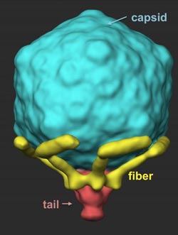
Researchers found that the T7 virus has six tail fibers that are folded back against its capsid. The fibers extend as the virus locates a suitable host and as it “walks” across its host cell surface to find a site to infect.<br><br>Credit: From Hu et. al. 2013. Science Express.<br>
To infect a cell, a virus must be able to first find a suitable cell and then eject its genetic material into its host. This robot-like process has been observed in a virus called T7 and visualized by Ian Molineux, professor of biology at The University of Texas at Austin, and his colleagues.
The researchers show that when searching for its prey, the virus briefly extends — like feelers — one or two of six ultra-thin fibers it normally keeps folded at the base of its head.
Once a suitable host has been located, the virus behaves a bit like a planetary rover, extending these fibers to walk randomly across the surface of the cell and find an optimal site for infection.
At the preferred infection site, the virus goes through a major change in structure in which it ejects some of its proteins through the bacterium's cell membrane, creating a path for the virus's genetic material to enter the host.
After the viral DNA has been ejected, the protein path collapses and the infected cell membrane reseals.
“Although many of these details are specific to T7,” said Molineux, “the overall process completely changes our understanding of how a virus infects a cell.”
For example, the researchers now know that most of the fibers are usually bound to the virus head rather than extended, as was previously thought. That those fibers are in a dynamic equilibrium between bound and extended states is also new.
Molineux said that the idea that phages “walk” over the cell surface was previously proposed, but their paper provides the first experimental evidence that this is the case.
This is also the first time that scientists have made actual images showing how the virus's tail extends into the host — the very action that allows it to infect a cell with its DNA.
“I first hypothesized that T7 made an extended tail more than 10 years ago,” said Molineux, “but this is the first irrefutable experimental evidence for the idea and provides the first images of what it looks like.”
The researchers used a combination of genetics and cryo-electron tomography to image the infection process. Cryo-electron tomography is a process similar to a CT scan, but it is scaled to study objects with a diameter a thousandth the thickness of a human hair.
Molineux's co-authors are Bo Hu, William Margolin and Jun Liu from UT Health.
Additional contacts:
Ian Molineux, Professor
The University of Texas at Austin
512-471-3143 molineux@austin.utexas.edu
Rob Cahill, Media Relations
UTHealth
713-500-3042
Robert.Cahill@uth.tmc.edu
Media Contact
More Information:
http://www.utexas.eduAll latest news from the category: Life Sciences and Chemistry
Articles and reports from the Life Sciences and chemistry area deal with applied and basic research into modern biology, chemistry and human medicine.
Valuable information can be found on a range of life sciences fields including bacteriology, biochemistry, bionics, bioinformatics, biophysics, biotechnology, genetics, geobotany, human biology, marine biology, microbiology, molecular biology, cellular biology, zoology, bioinorganic chemistry, microchemistry and environmental chemistry.


Newest articles

High-energy-density aqueous battery based on halogen multi-electron transfer
Traditional non-aqueous lithium-ion batteries have a high energy density, but their safety is compromised due to the flammable organic electrolytes they utilize. Aqueous batteries use water as the solvent for…

First-ever combined heart pump and pig kidney transplant
…gives new hope to patient with terminal illness. Surgeons at NYU Langone Health performed the first-ever combined mechanical heart pump and gene-edited pig kidney transplant surgery in a 54-year-old woman…

Biophysics: Testing how well biomarkers work
LMU researchers have developed a method to determine how reliably target proteins can be labeled using super-resolution fluorescence microscopy. Modern microscopy techniques make it possible to examine the inner workings…

