New protocol enables analysis of metabolic products from fixed tissues
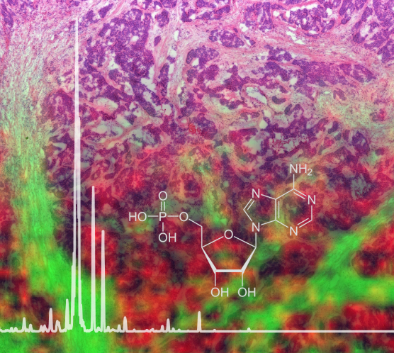
A new mass spectrometry imaging protocol allows the analysis of metabolites like Adenosine monophosphate from FFPE tissue, shown here as background. Source: Helmholtz Zentrum München
In biomedical research, working with tissue samples is indispensable because it permits insights into the biological reality of patients, for example, in addition to those gained from Petri dishes and computer simulations. The tissue is usually fixed in formalin and embedded in paraffin wax in order to keep the tissue, as far as possible, in its original condition for later analyses.
It was previously assumed that in material that had been treated in this way an analysis of metabolites, in contrast to DNA or proteins, would be barely possible for technical reasons. A team of scientists from the Analytical Pathology department at the Helmholtz Zentrum München led by Prof. Axel Karl Walch has now succeeded in refuting this belief.
Fixed tissues accessible on a large scale
The researchers developed a protocol which makes it possible – within one day – to determine the metabolite composition of tissues using a mass spectrometry imaging approach, and to make it visible in tissue sections. Relatively small amounts of material are required for this, according to the authors. “Our method permits the analysis of minute biopsies and even tissue micro-arrays, making it particularly interesting for molecular research and diagnostics,” explains doctoral candidate Achim Buck, together with Alice Ly, the first author of the study.
In order to ensure that the measured data was not falsified by the fixation process, the authors compared it with the measured values for the same samples that were not fixed but were shock frozen. “A large proportion of the measured metabolites occurred in both analyses,” reports Achim Buck. “We were able to show that the method works reliably and avoids the complex logistics and storage of shock-frozen samples.”
In addition to simple handling and high reproducibility, the possibility to conduct high throughput work is a key advantage of the new method*, according to the scientists. Above all, however, it is now possible to study the spatial distribution of molecules in the tissue graphically and with great precision. “That is an enormous advantage, both in research and in clinical diagnostic practice,” research team leader Walch says, assessing the new possibilities. “Using our new analytical method, our aim is now to identify new predictive, diagnostic and prognostic markers in tissues, as well as to understand disease processes.”
The scientists hope that publication of the protocol will also lead to an exchange with and further developments by colleagues with a view to advancing metabolic analyses of archived tissues.
Further Information
Background:
* Via tissue microarrays, which permit the analysis of several hundred patients in one measurement, tissue-based scientific and diagnostic questions relating to the pathogenesis of diseases and new treatment options can be clarified on a high throughput basis.
Original publication:
Ly, A. & Buck, A. et al. (2016). High Mass Resolution MALDI Mass Spectrometry Imaging of Metabolites from Formalin-Fixed Paraffin Embedded Tissue, Nature Protocols, DOI: nprot.2016.081
Supporting publication:
Buck, A. & Ly, A. et al. (2015). High-resolution MALDI-FT-ICR MS Imaging for the analysis of metabolites from formalin-fixed paraffin-embedded clinical tissue samples, The Journal of Pathology, doi:10.1002/path.4560
The Helmholtz Zentrum München, the German Research Center for Environmental Health, pursues the goal of developing personalized medical approaches for the prevention and therapy of major common diseases such as diabetes and lung diseases. To achieve this, it investigates the interaction of genetics, environmental factors and lifestyle. The Helmholtz Zentrum München is headquartered in Neuherberg in the north of Munich and has about 2,300 staff members. It is a member of the Helmholtz Association, a community of 18 scientific-technical and medical-biological research centers with a total of about 37,000 staff members. http://www.helmholtz-muenchen.de/en
The Analytical Pathology Department (AAP) carries out scientific development, as a complement to research units with a clinical and fundamental orientation, of translational research on diseases that occur in tissue. AAP is involved in the translation of (for example) in-vitro models or animal models to application in humans. AAP thus links, in collaboration with the Institute for Pathology (PATH), basic research with diagnostic application, subsequently translating the findings of experimental and molecular pathology into procedures for the classification of diseases and predictive diagnostics dealing with tissue. http://www.helmholtz-muenchen.de/aap
Contact for the media:
Department of Communication, Helmholtz Zentrum München – German Research Center for Environmental Health, Ingolstädter Landstr. 1, 85764 Neuherberg – Tel. +49 89 3187 2238 – Fax: +49 89 3187 3324 – E-mail: presse@helmholtz-muenchen.de
Scientific Contact at Helmholtz Zentrum München:
Prof. Dr. Axel Karl Walch, Helmholtz Zentrum München – German Research Center for Environmental Health, Analytical Pathology Department, Ingolstädter Landstr. 1, 85764 Neuherberg – Tel. +49 89 3187 2739, E-mail: axel.walch@helmholtz-muenchen.de
http://www.nature.com/nprot/journal/v11/n8/full/nprot.2016.081.html
Media Contact
All latest news from the category: Life Sciences and Chemistry
Articles and reports from the Life Sciences and chemistry area deal with applied and basic research into modern biology, chemistry and human medicine.
Valuable information can be found on a range of life sciences fields including bacteriology, biochemistry, bionics, bioinformatics, biophysics, biotechnology, genetics, geobotany, human biology, marine biology, microbiology, molecular biology, cellular biology, zoology, bioinorganic chemistry, microchemistry and environmental chemistry.


Newest articles

A universal framework for spatial biology
SpatialData is a freely accessible tool to unify and integrate data from different omics technologies accounting for spatial information, which can provide holistic insights into health and disease. Biological processes…

How complex biological processes arise
A $20 million grant from the U.S. National Science Foundation (NSF) will support the establishment and operation of the National Synthesis Center for Emergence in the Molecular and Cellular Sciences (NCEMS) at…

Airborne single-photon lidar system achieves high-resolution 3D imaging
Compact, low-power system opens doors for photon-efficient drone and satellite-based environmental monitoring and mapping. Researchers have developed a compact and lightweight single-photon airborne lidar system that can acquire high-resolution 3D…

