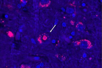
MRI scans in premature infants can predict future developmental delays
Terrie E. Inder, M.D., associate professor of pediatrics, of radiology and of neurology at Washington University School of Medicine in St. Louis, and pediatric researchers in New Zealand and Australia found that the magnetic resonance imaging (MRI) scans were able to determine abnormalities in the white matter and gray matter of the brains of very pre-term infants, those born at 30 weeks or less. Following the infants from birth to age 2, the researchers were able to grade those abnormalities to predict the risk of severe cognitive delays, psychomotor delays, cerebral palsy, or hearing or visual impairments that may be visible by age 2.
The results of the study appear in the Aug. 17 issue of the New England Journal of Medicine. The researchers studied 167 preterm infants in New Zealand and Australia and at St. Louis Children's Hospital. Inder said the findings are a breakthrough because previous technology — cranial ultrasounds — did not show the abnormalities in the infants' brains.
“With the MRI, now we can understand what's going wrong in the developing brain when the baby is born early,” Inder said. “We can use the MRI when the baby reaches full-term (40 weeks) to predict neurodevelopmental outcomes.” More than 2 percent of all live births are infants born before 32 weeks of gestation. Nationwide, the rate of premature births jumped 13 percent between 1992 and 2002, according to the March of Dimes. Recent data show that 50 percent of children born prematurely suffer some neurodevelopmental challenges, such as crawling, walking upright, running, swinging arms, and other activities that require coordination and balance. Among pre-term infants who survive, 5 percent to 15 percent have cerebral palsy, severe vision or hearing impairment or both, and 25 percent to 50 percent have cognitive, behavioral and social difficulties that require special educational resources.
The MRI scans show lesions on the infants' brains, as well as which region of the brain is affected and the severity of the risk for future developmental delays. For example, if a lesion is in the area of the brain that controls fine and gross motor skills, the risk is higher that the child will have some type of developmental delay in movement. Pediatricians would then know that the child would benefit from immediate physical therapy, Inder said.
“We can use these results to determine which baby would benefit most from physical, occupational or speech therapy,” Inder said. “We can also help prepare the parents for future challenges with learning delays and developmental disabilities.”
Media Contact
More Information:
http://www.wustl.eduAll latest news from the category: Health and Medicine
This subject area encompasses research and studies in the field of human medicine.
Among the wide-ranging list of topics covered here are anesthesiology, anatomy, surgery, human genetics, hygiene and environmental medicine, internal medicine, neurology, pharmacology, physiology, urology and dental medicine.


Newest articles
After 25 years, researchers uncover genetic cause of rare neurological disease
Some families call it a trial of faith. Others just call it a curse. The progressive neurological disease known as spinocerebellar ataxia 4 (SCA4) is a rare condition, but its…
Lower dose of mpox vaccine is safe
… and generates six-week antibody response equivalent to standard regimen. Study highlights need for defined markers of mpox immunity to inform public health use. A dose-sparing intradermal mpox vaccination regimen…
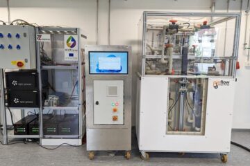
Efficient, sustainable and cost-effective hybrid energy storage system for modern power grids
EU project HyFlow: Over three years of research, the consortium of the EU project HyFlow has successfully developed a highly efficient, sustainable, and cost-effective hybrid energy storage system (HESS) that…

