Brain volume and memory impairment
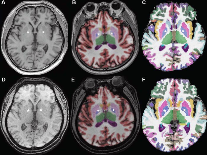
Conventional 3D T1W image (A) without segmentation, (B) with segmentation by Neuroquant, and (C) with segmentation by Freesurfer; ultrafast 3D-EPI T1W image (D) without segmentation, (E) with segmentation by Neuroquant, and (F) with segmentation by Freesurfer. Pallidum (asterisks) appears larger for Freesurfer (C, F) than for NeuroQuant (B, E), but appears similar in size between two sequences for each software package. For both software packages, bilateral frontal and occipital cortices (B, C) at bone-tissue interface appear more color-coded for conventional than for ultrafast sequence, contributing to larger cortical gray matter for conventional sequence.
Credit: American Roentgen Ray Society (ARRS), American Journal of Roentgenology (AJR)
… conventional vs ultrafast 3D MRI sequences.
Automated brain volumetry in memory-impaired patients shows significant differences and systematic biases between conventional and ultrafast 3D T1-weighted MRI sequences.
According to an article in ARRS’ American Journal of Roentgenology (AJR), brain volume measurements in memory-impaired patients show significant differences and systematic biases between conventional and ultrafast 3D T1-weighted (T1W) MRI sequences.
“In patients for whom severe motion artifact precludes use of the conventional sequence, the ultrafast sequence may be useful to enable brain volumetry,” wrote corresponding author Hye Jin Baek of Gyeongsang National University and Changwon Hospital in Korea. “However, the current conventional 3D T1W sequence remains preferred in patients who can tolerate the standard examination.”
To compare automated brain volume measurements between conventional 3D T1W and ultrafast 3D echo-planar imaging (EPI) T1W sequences, Baek and colleagues retrospectively studied 36 patients (25 women, 11 men; mean age, 68 years) with memory impairment who underwent 3-T brain MRI. Examinations included both isotropic 3D T1W using inversion recovery gradient-recalled echo sequence (slice, 1.0 mm; acquisition, 3:04) and, in patients exhibiting motion, isotropic 3D EPI T1W sequence (slice, 1.2 mm; acquisition, 0:30). Using NeuroQuant (CorTechs Labs) and FreeSurfer (Harvard University) software to automate brain segmentation, measurements were compared between sequences for nine regions in each hemisphere.
For automated brain volumetry from ultrafast 3D EPI T1W imaging, compared with conventional 3D T1W imaging, most regions demonstrated at least substantial agreement between the two sequences—yet also significantly different mean values, moderate or large effect sizes, and consistent systematic biases with wide limits of agreement.
“The variation between the two sequences was observed in subset analyses of 16 patients with, and 20 patients without, Alzheimer disease,” the authors of this AJR article added.
An electronic supplement to this AJR article is available here.
Founded in 1900, the American Roentgen Ray Society (ARRS) is the first and oldest radiological society in North America, dedicated to the advancement of medicine through the profession of radiology and its allied sciences. An international forum for progress in medical imaging since the discovery of the x-ray, ARRS maintains its mission of improving health through a community committed to advancing knowledge and skills with an annual scientific meeting, monthly publication of the peer-reviewed American Journal of Roentgenology (AJR), quarterly issues of InPractice magazine, AJR Live Webinars and Podcasts, topical symposia, print and online educational materials, as well as awarding scholarships via The Roentgen Fund®.
MEDIA CONTACT:
Logan K. Young, PIO
44211 Slatestone Court
Leesburg, VA 20176
703-858-4332
Journal: American Journal of Roentgenology
DOI: 10.2214/AJR.21.27043
Method of Research: Observational study
Subject of Research: People
Article Title: Automated Brain Volumetry in Patients With Memory Impairment: Comparison of Conventional and Ultrafast 3D T1-weighted MRI Sequences Using Two Software Packages
Article Publication Date: 5-Jan-2022
COI Statement: N/A
Media Contact
Logan Young
American Roentgen Ray Society
lyoung@arrs.org
Office: 703-858-4332
All latest news from the category: Medical Engineering
The development of medical equipment, products and technical procedures is characterized by high research and development costs in a variety of fields related to the study of human medicine.
innovations-report provides informative and stimulating reports and articles on topics ranging from imaging processes, cell and tissue techniques, optical techniques, implants, orthopedic aids, clinical and medical office equipment, dialysis systems and x-ray/radiation monitoring devices to endoscopy, ultrasound, surgical techniques, and dental materials.


Newest articles

High-energy-density aqueous battery based on halogen multi-electron transfer
Traditional non-aqueous lithium-ion batteries have a high energy density, but their safety is compromised due to the flammable organic electrolytes they utilize. Aqueous batteries use water as the solvent for…

First-ever combined heart pump and pig kidney transplant
…gives new hope to patient with terminal illness. Surgeons at NYU Langone Health performed the first-ever combined mechanical heart pump and gene-edited pig kidney transplant surgery in a 54-year-old woman…

Biophysics: Testing how well biomarkers work
LMU researchers have developed a method to determine how reliably target proteins can be labeled using super-resolution fluorescence microscopy. Modern microscopy techniques make it possible to examine the inner workings…

