New insights into lung tissue in COVID-19 disease

Sections through the three-dimensional reconstruction volume (upper left, grey) around a pulmonary alveolus with hyaline membrane (lower left, yellow). On the right, the images are superimposed. In the centre is the air bubble (alveolus). The electron density is represented by different shades of grey. On the inside of the air bubble is a layer of proteins and dead cell residues, the "hyaline membrane". This deposit, which can be represented in its three-dimensional structure for the first time by the new method, reduces the gas exchange and leads to respiratory distress.
Credit: T Salditt, M Eckermann
Researchers led by Göttingen University develop new three-dimensional imaging technique to visualize tissue damage in severe Covid-19
Physicists at the University of Göttingen, together with pathologists and lung specialists at the Medical University of Hannover, have developed a three-dimensional imaging technique that enables high resolution and three-dimensional representation of damaged lung tissue following severe Covid-19.
Using a special X-ray microscopy technique, they were able to image changes caused by the coronavirus in the structure of alveoli (the tiny air sacs in the lung) and the vasculature. The results of the study were published in the research journal eLife.
In severe Covid-19 disease, the researchers observed significant changes in the vasculature, inflammation, blood clots and “hyaline membranes”, which are composed of proteins and dead cells deposited on the alveolar walls, which make gas exchange difficult or impossible. With their new imaging approach, these changes can be visualized for the first time in larger tissue volumes, without cutting and staining or damaging the tissue as in conventional histology.
It is particularly well suited for tracing small blood vessels and their branches in three dimensions, localizing cells of the immune systems which are recruited to the inflammation sites, and measuring the thickness of the alveolar walls. Due to the three-dimensional reconstruction, the data could also be used to simulate gas exchange.
“Using zoom tomography, large areas of lung tissue embedded in wax can be scanned enabling detailed examination to locate particularly interesting areas around inflammation, blood vessels or bronchial tubes,” says lead author Professor Tim Salditt from the Institute of X-ray Physics at the University of Göttingen. Since X-rays penetrate deep into tissue, this enables scientists to understand the relation between the microscopic tissue structure and the larger functional architecture of an organ. This is important, for example, to visualize the tree of blood vessels down to the smallest capillaries.
The authors foresee that this new X-ray technique will be an extension to traditional histology and histopathology, areas of study which go back to the 19th century when optical microscopes had just become available and pathologists could thereby unravel the microscopic origins of many diseases. Even today, pathologists still follow the same basic steps to prepare and investigate tissue: chemical fixation, slicing, staining and microscopy. This traditional approach, however, is not sufficient if three-dimensional images are required or if large volumes have to be screened, digitalized or analysed with computer programmes.
Three-dimensional imaging is well known from medical computerized tomography (CT). However, the resolution and contrast of this conventional technique are not sufficient to detect the tissue structure with cellular or sub-cellular resolution. Therefore, the authors used “phase contrast”, which exploits the different propagation velocities of X-rays in tissue to generate an intensity pattern on the detector.
Salditt and his research group at the Institute for X-ray Physics developed special illumination optics and algorithms to reconstruct sharp images from these patterns, an approach which they have now adapted for the study of lung tissue affected by severe progression of Covid-19. The Göttingen team could record lung tissue at scalable size and resolution, yielding both larger overviews and close-up reconstructions.
Depending on the setting, their method can even yield structural details below the resolution of conventional light microscopy. To achieve this, the researchers used highly powerful X-ray radiation generated at the PETRAIII storage ring of the German Electron Synchrotron (DESY) in Hamburg.
As was the case when the modern microscope was invented 150 years ago, significant progress has resulted from collaboration between physicists and medical researchers. The interdisciplinary research team hopes that the new method will support the development of treatment methods, medicines to prevent or alleviate severe lung damage in Covid-19, or to promote regeneration and recovery.
“It is only when we can clearly see and understand what is really going on, that we can develop targeted interventions and drugs,” adds Danny Jonigk (Medical University Hannover), who led the medical part of the interdisciplinary study.
###
Original publication: M. Eckermann, J.Frohn, M. Reichardt, M. Osterhoff, M. Sprung, F. Westermeier, A. Tzankov, C. Werlein, M. Kühnel, D. Jonigk, T. Salditt et al. “3d Virtual Patho-Histology of Lung Tissue from Covid-19 Patients based on Phase Contrast X-ray Tomography” eLife (2020). DOI: 10.7554/eLife.60408 (content also available on medRxiv: rhttps:/
Contact:
Professor Tim Salditt
University of Göttingen
Institute for X-ray Physics
Friedrich-Hund-Platz 1, 37077 Göttingen
Tel: +49 (0) 551 39 29918 / 25556
Email: tsaldit@gwdg.de
http://www.
Media Contact
All latest news from the category: Medical Engineering
The development of medical equipment, products and technical procedures is characterized by high research and development costs in a variety of fields related to the study of human medicine.
innovations-report provides informative and stimulating reports and articles on topics ranging from imaging processes, cell and tissue techniques, optical techniques, implants, orthopedic aids, clinical and medical office equipment, dialysis systems and x-ray/radiation monitoring devices to endoscopy, ultrasound, surgical techniques, and dental materials.


Newest articles
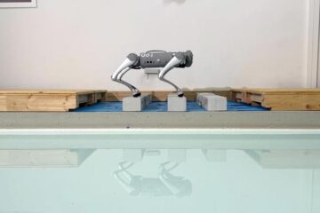
Trotting robots reveal emergence of animal gait transitions
A four-legged robot trained with machine learning by EPFL researchers has learned to avoid falls by spontaneously switching between walking, trotting, and pronking – a milestone for roboticists as well…
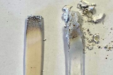
Innovation promises to prevent power pole-top fires
Engineers in Australia have found a new way to make power-pole insulators resistant to fire and electrical sparking, promising to prevent dangerous pole-top fires and reduce blackouts. Pole-top fires pose…
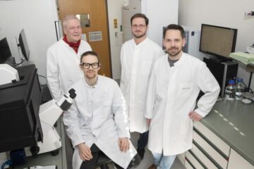
Possible alternative to antibiotics produced by bacteria
Antibacterial substance from staphylococci discovered with new mechanism of action against natural competitors. Many bacteria produce substances to gain an advantage over competitors in their highly competitive natural environment. Researchers…

