Bye, bye, biopsy?

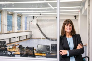
A handheld device being developed at Stevens Institute of Technology can scan skin and detect different types of skin cancer, such as carcinoma (left), squamous cell carcinoma (middle), and actinic keratosis (right).
Credit: Stevens Institute of Technology
Handheld device could painlessly identify skin cancers.
Stevens Institute of Technology uses millimeter-wave imaging to slash rate of unnecessary biopsies.
Skin biopsies are no fun: doctors carve away small lumps of tissue for laboratory testing, leaving patients with painful wounds that can take weeks to heal. That’s a price worth paying if it enables early cancer treatment. However, in recent years, aggressive diagnostic efforts have seen the number of biopsies grow around four times faster than the number of cancers detected, with about 30 benign lesions now biopsied for every case of skin cancer that’s found.
Researchers at Stevens Institute of Technology are now developing a low-cost handheld device that could cut the rate of unnecessary biopsies in half and give dermatologists and other frontline physicians easy access to laboratory-grade cancer diagnostics. “We aren’t trying to get rid of biopsies,” said Negar Tavassolian, director of the Bio-Electromagnetics Laboratory at Stevens. “But we do want to give doctors additional tools and help them to make better decisions.”
The team’s device uses millimeter-wave imaging — the same technology used in airport security scanners — to scan a patient’s skin. (In earlier work, Tavassolian and her team had to work with already biopsied skin for the device to detect if it was cancerous.)
Healthy tissue reflects millimeter-wave rays differently than cancerous tissue, so it’s theoretically possible to spot cancers by monitoring contrasts in the rays reflected back from the skin. To bring that approach into clinical practice, the researchers used algorithms to fuse signals captured by multiple different antennas into a single ultrahigh-bandwidth image, reducing noise and quickly capturing high-resolution images of even the tiniest mole or blemish.
Spearheaded by Amir Mirbeik Ph.D. ’18, the team used a tabletop version of their technology to examine 71 patients during real-world clinical visits, and found their methods could accurately distinguish benign and malignant lesions in just a few seconds. Using their device, Tavassolian and Mirbeik could identify cancerous tissue with 97% sensitivity and 98% specificity — a rate competitive with even the best hospital-grade diagnostic tools.
“There are other advanced imaging technologies that can detect skin cancers, but they’re big, expensive machines that aren’t available in the clinic,” said Tavassolian, whose work appears in the March 23 issue of Scientific Reports. “We’re creating a low-cost device that’s as small and as easy to use as a cellphone, so we can bring advanced diagnostics within reach for everyone.”
Because the team’s technology delivers results in seconds, it could one day be used instead of a magnifying dermatoscope in routine checkups, giving extremely accurate results almost instantly. “That means doctors can integrate accurate diagnostics into routine checkups, and ultimately treat more patients,” said Tavassolian.
Unlike many other imaging methods, millimeter-wave rays harmlessly penetrate about 2mm into human skin, so the team’s imaging technology provides a clear 3D map of scanned lesions. Future improvements to the algorithm powering the device could significantly improve mapping of lesion margins, enabling more precise and less invasive biopsying for malignant lesions.
The next step is to pack the team’s diagnostic kit onto an integrated circuit, a step that could soon allow functional handheld millimeter-wave diagnostic devices to be produced for as little as $100 a piece — a fraction of the cost of existing hospital-grade diagnostic equipment. The team is already working to commercialize their technology and hopes to start putting their devices in clinicians’ hands within the next two years.
“The path forward is clear, and we know what we need to do,” said Tavassolian. “After this proof of concept, we need to miniaturize our technology, bring the price down, and bring it to the market.”
Journal: Scientific Reports
DOI: 10.1038/s41598-022-09047-6
Method of Research: Experimental study
Subject of Research: People
Article Title: Real‑time high‑resolution millimeter‑wave imaging for in‑vivo skin cancer diagnosis
Article Publication Date: 23-Mar-2022
Media Contact
Thania Benios
Stevens Institute of Technology
thania.benios@stevens.edu
Office: 917-930-5988
All latest news from the category: Medical Engineering
The development of medical equipment, products and technical procedures is characterized by high research and development costs in a variety of fields related to the study of human medicine.
innovations-report provides informative and stimulating reports and articles on topics ranging from imaging processes, cell and tissue techniques, optical techniques, implants, orthopedic aids, clinical and medical office equipment, dialysis systems and x-ray/radiation monitoring devices to endoscopy, ultrasound, surgical techniques, and dental materials.


Newest articles
Combining robotics and ChatGPT
TUM professor uses ChatGPT for choreographies with flying robots. Prof. Angela Schoellig has proved that large language models can be used safely in robotics. ChatGPT develops choreographies for up to…

How the Immune System Learns from Harmless Particles
Our lungs are bombarded by all manner of different particles every single day. Whilst some are perfectly safe for us, others—known as pathogens—have the potential to make us ill. The…

Biomarkers identified for successful treatment of bone marrow tumours
CAR T cell therapy has proven effective in treating various haematological cancers. However, not all patients respond equally well to treatment. In a recent clinical study, researchers from the University…

