The metabolism of bacteria

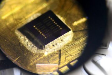
Metabolites in microbiomes on the micron scale. Each color in the picture represents a different metabolite, imaged with Mass spectrometry imaging at highest pixel resolution (2 um). The three yellow hotspots show epithel cells infected with a bacterium and its metabolic consequences.
© Borceau et al. Nature Protocols 2023
New method reveals host-microbe interactions.
Joint press release by the CAU and the Max Planck Institute for Marine Microbiology: North German researchers are developing a method that reveals the chemical communication between microbes and their host.
The fascinating world of bacteria that live as symbionts or parasites in animal hosts often remains a mystery to researchers. Kiel University (CAU) and the Max Planck Institute for Marine Microbiology in Bremen are contributing to solving this puzzle with their research into the interactions between microbes and their host. Under the leadership of Prof. Dr Manuel Liebeke, head of the Metabolomics Department at the CAU’s Faculty of Agricultural and Nutritional Sciences and the Metabolic Interactions Research Group at the Max Planck Institute for Marine Microbiology, a research team has made a breakthrough that provides insights into this mysterious micro-world.
Often, bacteria cannot be cultured in the laboratory, and researchers must rely on information from the bacterial genome obtained from environmental samples to gain theoretical insights into the metabolism of microorganisms. However, there has been a lack of insight into what they actually do in their natural environment. To solve this puzzle, scientists began researching the so-called metabolome of bacteria – everything that has to do with their metabolism, including metabolites such as sugars or fats.
In a pioneering study, Liebeke’s team developed a method with which they can identify individual bacteria and simultaneously determine which metabolites are present in the cells – all without cultivating the bacteria in the laboratory. This method allows them to study how bacteria live and survive as symbiotic subtenants, for example in mussels. The team analysed hundreds of metabolites on an area smaller than one square millimetre. The Kiel and Bremen researchers published their results in the journal Nature Protocols in September.
A frozen moment enables detailed observation
“We create a snapshot, so to speak, of the bacteria at work, exactly as they are active in their natural environment, particularly within an animal cell,” Liebeke explained. “And we can do that at an impressive resolution of a few micrometres, about ten times thinner than a human hair.”
A special feature of this method is the use of flash-frozen tissue, which is cut wafer-thin. The researchers then use a special mass spectrometry technique called MALDI-MS imaging to create a snapshot of the chemical compounds in the cells.
However, drawing the correct conclusions from the images of the metabolites is only possible if they know which bacteria produce or use them. To solve this problem, the researchers also use fluorescence in situ hybridisation (FISH) to identify and localise individual bacterial cells in the sample.
“Applying this method to host-microbe communities will give us many exciting new insights into chemical communication between organisms,” said Patric Bourceau from the Max Planck Institute for Marine Microbiology, lead author of the protocol developed to apply the method.
This groundbreaking work opens new doors for the study of bacteria and their interactions with their host. In addition, the method presented here also offers promising potential applications for the future: developed at the Max Planck Institute in Bremen, Liebeke’s new working group at the CAU is now using it to study the human gut microbiome and its influence on metabolism. This could, for example, help us to better understand inflammatory bowel diseases. With the publication of a detailed protocol, the application of the technique is now open to other researchers worldwide.
In summary, the application of microscopy and metabolomics (the name of the research field dedicated to the study of metabolites) provides insights into the functional and chemical ecology of host-microbe interactions. The steady advances in MALDI-MSI technology make it possible to illustrate microbial colonies, biofilms and individual eukaryotic cells, and even bacterial microcolonies. Today, MALDI-MSI technology is on the verge of being able to provide images of individual bacterial cells. The protocol presented here forms the basis for analysing and understanding metabolic interactions, down to the micrometre.
Photos are available for download at:
http://www.uni-kiel.de/de/pressemitteilungen/2023/270-stoffwechsel-bakterien-liebeke.jpg
Prof. Dr Manuel Liebeke, head of the Metabolomics Department at the CAU’s Faculty of Agricultural and Nutritional Sciences and the Metabolic Interactions Research Group at the Max Planck Institute for Marine Microbiology.
© Fanni Aspetsberger, Max Planck Institute for Marine Microbiology
http://www.uni-kiel.de/de/pressemitteilungen/2023/270-stoffwechsel-bakterien-bourceau.jpeg
Patric Bourceau from the Max Planck Institute for Marine Microbiology, lead author of the protocol.
© Manuel Liebeke, Kiel University
http://www.uni-kiel.de/de/pressemitteilungen/2023/270-MPI-CAU.jpg
Metabolites in microbiomes on the micron scale. Each color in the picture represents a different metabolite, imaged with Mass spectrometry imaging at highest pixel resolution (2 um). The three yellow hotspots show epithel cells infected with a bacterium and its metabolic consequences.
© Borceau et al. Nature Protocols 2023
Kiel University
Press, Communication and Marketing, Eva Sittig, Text/editing: Prof. Dr Manuel Liebeke
Postal address: D-24098 Kiel, Germany, Telephone: (0431) 880-2104, Fax: (0431) 880-1355
E-mail: presse@uv.uni-kiel.de, Internet: http://www.uni-kiel.de Twitter: http://www.twitter.com/kieluni
Facebook: http://www.facebook.com/kieluni Instagram: http://www.instagram.com/kieluni
Wissenschaftliche Ansprechpartner:
Prof. Dr Manuel Liebeke
Kiel University
Institute of Human Nutrition and Food Science
Metabolomics Department
mliebeke@metabolomics.uni-kiel.de
Originalpublikation:
https://www.nature.com/articles/s41596-023-00864-1
Patric Bourceau, Benedikt Geier, Vincent Suerdieck, Tanja Bien, Jens Soltwisch, Klaus Dreisewerd & Manuel Liebeke (2023): Visualization of metabolites and microbes at high spatial resolution using MALDI mass spectrometry imaging and in situ fluorescence labeling.
Nature Protocols
First published: 06.09.2023 DOI: 10.1038/s41596-023-00864-1
Weitere Informationen:
http://www.uni-kiel.de/en/details/news/en-270-metabolism-bacteria
Media Contact
All latest news from the category: Life Sciences and Chemistry
Articles and reports from the Life Sciences and chemistry area deal with applied and basic research into modern biology, chemistry and human medicine.
Valuable information can be found on a range of life sciences fields including bacteriology, biochemistry, bionics, bioinformatics, biophysics, biotechnology, genetics, geobotany, human biology, marine biology, microbiology, molecular biology, cellular biology, zoology, bioinorganic chemistry, microchemistry and environmental chemistry.


Newest articles

Sea slugs inspire highly stretchable biomedical sensor
USC Viterbi School of Engineering researcher Hangbo Zhao presents findings on highly stretchable and customizable microneedles for application in fields including neuroscience, tissue engineering, and wearable bioelectronics. The revolution in…

Twisting and binding matter waves with photons in a cavity
Precisely measuring the energy states of individual atoms has been a historical challenge for physicists due to atomic recoil. When an atom interacts with a photon, the atom “recoils” in…
Nanotubes, nanoparticles, and antibodies detect tiny amounts of fentanyl
New sensor is six orders of magnitude more sensitive than the next best thing. A research team at Pitt led by Alexander Star, a chemistry professor in the Kenneth P. Dietrich…

