SARS-CoV-2 infects sustentacular cells in the olfactory epithelium of COVID-19 patients
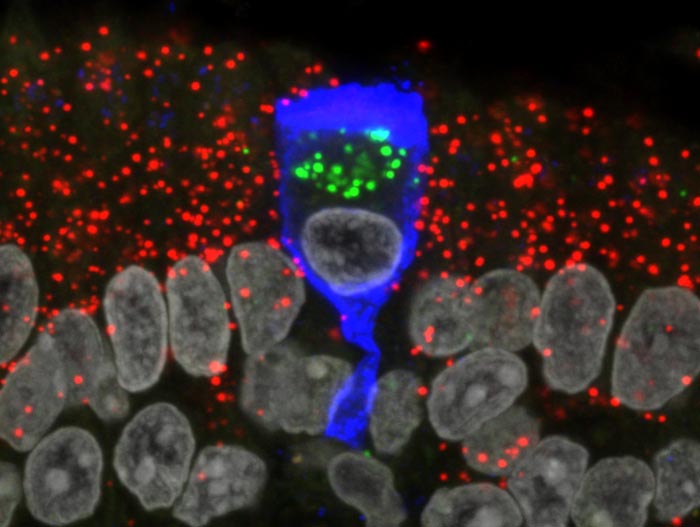
A lone infected sustentacular cell is surrounded by non-infected cells in the olfactory mucosa of a COVID-19 patient who died four days after diagnosis of the infection. The infected cell has the characteristic shape of a wine glass. The blue color comes from staining with an antibody against the nucleocapsid protein of the virus. The red dots represent staining with an RNAscope probe for a gene that is expressed in sustentacular cells (GPX3). Within the lone infected cell, there are few or no red dots, because infection of a cell with SARS-CoV-2 causes decay of host RNA molecules. The green dots represent staining with an an RNAscope probe for a type of viral RNA molecules that are only present during ongoing viral replication. This lone sustentacular cell was thus serving as a “factory” for replicating viral RNA at the time the postmortem tissue sample was taken.
Credit: MaxPlanck Research Unit f. Neurogenetics/ Mona Khan
The coronavirus does not appear to infect nerve cells in the olfactory epithelium and in the olfactory bulb.
It is now widely known that COVID-19 is associated with the transient or long-term loss of olfaction (the sense of smell) but the mechanisms remain obscure. An unresolved question is whether the olfactory nerve can provide SARS-CoV-2 with a route of entry to the brain. Scientists at the Max Planck Research Unit for Neurogenetics in Frankfurt in collaboration with physicians and scientists at the University Hospitals Leuven (Leuven, Belgium) and a major hospital in Bruges, Belgium, together with scientists at NanoString Technologies Inc. in Seattle, USA, report that SARS-CoV-2 does not appear to infect the sensory neurons of the olfactory epithelium in COVID-19 patients. Moreover, the team failed to find evidence for infection of olfactory bulb neurons. Instead, the sustentacular cells, also known as supporting cells, are the main target cell type for the virus in the olfactory epithelium. Since SARS-CoV2 spares olfactory sensory neurons and olfactory bulb neurons, it does not appear to be a neurotropic virus.
To infect a cell, SARS-CoV-2 must bind to a receptor on the cell membrane, and the classic entry receptor is ACE2. Earlier studies had shown that ACE2 is expressed by sustentacular cells in the human olfactory epithelium but not by olfactory sensory neurons, the nerve cells that are stimulated by odorants in the inhaled air and that transmit electrical signals to the olfactory bulb. There is no literature about the functions of sustentacular cells in the olfactory epithelium of humans. Studies in laboratory animals suggest that sustentacular cells provide olfactory sensory neurons with a variety of supportive functions, including structural and metabolic support. Both cell types are continuously regenerated from stem cells within the olfactory epithelium throughout the life of an individual.
As the olfactory mucosa is hidden deep within the nasal cavity, tissue sample procurement is not a practical option in patients while they are suffering from COVID-19. Hence, the physicians developed a novel protocol for harvesting tissue samples from deceased COVID-19 patients. As a control, tissue samples were taken from patients who had died from other causes and who were not infected with SARS-CoV-2 at the time of death. The workflow started with the notification of a team of ear, nose, & throat physicians about the death of a COVID-19 patient in an intensive care unit or ward. Using an endoscope, the physicians collected samples from the respiratory and olfactory mucosae and both olfactory bulbs. They were able to do so within 60 to 90 minutes after the death of the patient. “Thanks to this short postmortem interval, the tissue samples were in pristine condition for molecular biology studies,” says Laura Van Gerven, an ear, nose, & throat surgeon in Leuven and co-principal investigator of the project called ANOSMIC-19.
Analysis using RNAscope
The team of scientists in Frankfurt was led by Mona Khan. They used specially designed probes to stain sections of the tissue samples and analyze them under a confocal microscope. The ultrasensitive analytical method, also known as RNAscope, makes it possible to visualize various types of RNA molecules of SARS-CoV-2 within single cells. The scientists were able to assign the infected cells to specific cell types by simultaneously visualizing, in distinct colors, RNA molecules that are characteristic of various cell types, in combination with classical cell staining methods using antibodies. “Our results show that SARS-CoV-2 infects sustentacular cells in the olfactory epithelium of COVID-19 patients and replicates vigorously within these cells,” says Peter Mombaerts, director of the Max Planck Research Unit for Neurogenetics.
Applying a novel approach of whole-transcriptome analysis using Digital Spatial Profiler from NanoString Technologies Inc., analysis of sections of the olfactory mucosa of a COVID-19 patient revealed that infection of sustentacular cells does not alter the expression of olfactory receptor genes in nearby olfactory sensory neurons.
Viral RNA in the leptomeninges
Viral RNA could not be detected in olfactory bulb neurons either. Interestingly, in a third of cases, the researchers detected viral RNA in the meninges surrounding the olfactory bulb, the so-called leptomeninges. In these anatomical locations, the viral RNA may not be present in cells that had been infected with the virus but may stem from virus particles that may have entered the leptomeninges by hitchhiking on the olfactory nerve or via the blood stream. Alternatively, the viral RNA in the leptomeninges may simply represent viral RNA molecules that were floating around in the blood and not packaged in viral particles.
Thus, the results do not support previous suggestions that SARS-CoV-2 can infect nerve cells in humans. In other words, SARS-CoV-2 does not appear to be a neurotropic virus. The multidisciplinary team postulates that transient olfactory dysfunction in COVID-19 is triggered ty transient insufficient support from sustentacular cells to olfactory sensory neurons. The virus would thus affect olfactory sensory neurons indirectly but without infecting them directly. The pathological consequences of infection of sustentacular cells could vary from patient to patient. The researchers speculate that the immune system may be unable to provide sustentacular cells with full protection from infection, due to their location at the surface of the nasal mucosa. They further speculate that some vaccinated or recovered patients may still lose their sense of smell after exposure to SARS-CoV-2.
Journal: Cell
DOI: 10.1016/j.cell.2021.10.027
Method of Research: Experimental study
Article Title: Visualizing in deceased COVID-19 patients how SARS-CoV-2 attacks the respiratory and olfactory mucosae but spares the olfactory bulb.
Article Publication Date: 24-Nov-2021
All latest news from the category: Life Sciences and Chemistry
Articles and reports from the Life Sciences and chemistry area deal with applied and basic research into modern biology, chemistry and human medicine.
Valuable information can be found on a range of life sciences fields including bacteriology, biochemistry, bionics, bioinformatics, biophysics, biotechnology, genetics, geobotany, human biology, marine biology, microbiology, molecular biology, cellular biology, zoology, bioinorganic chemistry, microchemistry and environmental chemistry.


Newest articles

Sea slugs inspire highly stretchable biomedical sensor
USC Viterbi School of Engineering researcher Hangbo Zhao presents findings on highly stretchable and customizable microneedles for application in fields including neuroscience, tissue engineering, and wearable bioelectronics. The revolution in…

Twisting and binding matter waves with photons in a cavity
Precisely measuring the energy states of individual atoms has been a historical challenge for physicists due to atomic recoil. When an atom interacts with a photon, the atom “recoils” in…
Nanotubes, nanoparticles, and antibodies detect tiny amounts of fentanyl
New sensor is six orders of magnitude more sensitive than the next best thing. A research team at Pitt led by Alexander Star, a chemistry professor in the Kenneth P. Dietrich…

