Researchers detect a diffusion barrier inside fly brain
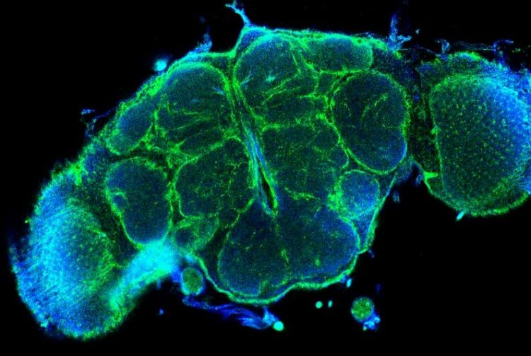
Optical cross-section through a fly brain. Green: distribution of the extracellular matrix protein "Trol", which is formed by all enveloping glial cells. The subdivision of the brain into different reaction spaces becomes clear.
(c) WWU - Nicole Pogodalla und Christian Klämbt
A team of researchers led by Nicole Pogodalla and Prof. Dr. Christian Klämbt from the Institute of Neuro- and Behavioral Biology at University of Münster now demonstrated for the first time an internal diffusion barrier in the brain of fruit flies – in addition to the already known blood-brain barrier.
The neurons, located in the brain are interconnected in a complex pattern and establish special communication points, the synapses. All neurons require a constant environment in order to function reliably. To ensure this, the brain is surrounded by the so-called blood-brain barrier. It ensures, for example, that the nutrient balance always remains the same and that harmful influences do not reach the neurons.
This applies to all animals including humans. For insects, a team led by Nicole Pogodalla and Prof. Dr. Christian Klämbt from the Institute of Neuro- and Behavioral Biology at the University of Münster has now shown that there is also a second barrier in the brain. Here glial cells, too, ensure a spatial separation of different functional compartments, which is essential for reliable functioning of the nervous system. The work was published in the prestigious online journal Nature Communications.
The research team studied the insect brain using larvae of the fruit fly (Drosophila melanogaster) as an example and focused on the role of glial cells. Early in development these cells help to establish the correct neuronal network and later glial cells play important roles in controlling the transmission of signals between neurons. In all invertebrates, as well as in primitive vertebrates, glial cells also define the outer boundary of the nervous system – the blood-brain barrier.
Deep in the fly brain, all synapses are located in a special region called the neuropil. The neuropil is separated from the zone containing the cell bodies of the neurons by a small set of surrounding glial cells, that were in the focus of Nicole Pogodalla. She developed a new experimental approach – dye injections into living larval brains – and combined this with cell type specific ablation experiments to show that these glial cells actually form a diffusion barrier, i. e. regulate the distribution of molecules.
Since all other cellular barriers in the body are formed by polarized cells that have an “up” and a “down,” the research team next examined glial cell polarity. Using advanced confocal image analysis as well as electron microscopy work in combination with state-of-the-art molecular genetics, the researchers uncovered that the ensheathing glial cells are indeed polarized. They showed that this polarization is functionally important, as defects in polarity lead to both an altered cell shape and cause a significant behavioral phenotype in fly larvae: Movement of larvae with defective or absent glial cells is impaired, and crawling speed is reduced.
In the current paper, the research team also describes the importance of extracellular matrix – the tissue lying between cells -, membrane lipids and membrane proteins, as well as the function of the cytoskeleton in the formation of the barrier-forming glial cells. The study includes extensive electron microscopy data obtained in a collaboration with Dr. Albert Cardona at the HHMI Janelia Research Campus in Ashburn, VA 20147, USA.
The German Research Foundation provided financial support for the work as part of Collaborative Research Center (SFB) 1348.
Wissenschaftliche Ansprechpartner:
Nicole Pogodalla
University of Münster
Institute of Neuro- and Behavioral Biology
Email: pogodall@uni-muenster.de
Originalpublikation:
Nicole Pogodalla, Holger Kranenburg, Simone Rey, Silke Rodrigues, Albert Cardona & Christian Klämbt (2021): Drosophila ßHeavy-Spectrin is required in polarized ensheathing glia that form a diffusion-barrier around the neuropil. Nature Communications, DOI:10.1038/s41467-021-26462-x
AG Klämbt: http://neurobio.uni-muenster.de/team-members
https://www.uni-muenster.de/news/view.php?cmdid=12138&lang=en
Media Contact
All latest news from the category: Life Sciences and Chemistry
Articles and reports from the Life Sciences and chemistry area deal with applied and basic research into modern biology, chemistry and human medicine.
Valuable information can be found on a range of life sciences fields including bacteriology, biochemistry, bionics, bioinformatics, biophysics, biotechnology, genetics, geobotany, human biology, marine biology, microbiology, molecular biology, cellular biology, zoology, bioinorganic chemistry, microchemistry and environmental chemistry.


Newest articles
Economies take off with new airports
A global study by an SUTD researcher in collaboration with scientists from Japan explores the economic benefits of airport investment in emerging economies using nighttime satellite imagery. Be it for…
CAR T–cell immunotherapy targets
Pan-cancer analysis uncovers a new class of promising CAR T–cell immunotherapy targets. Scientists at St. Jude Children’s Research Hospital found 156 potential CAR targets across the brain and solid tumors,…
Stony coral tissue loss disease
… is shifting the ecological balance of Caribbean reefs. The outbreak of a deadly disease called stony coral tissue loss disease is destroying susceptible species of coral in the Caribbean…

