Detailed image of the human retina
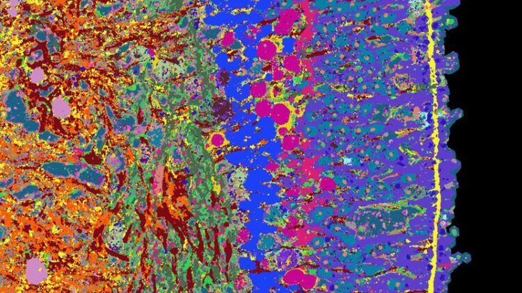
Detail of a cross-section of a retinal organoid. Different tissue structures are made visible with different colours.
Credit: Wahle et al. Nature Biotechnology 2023
What cell types are found in which human tissue, and where? Which genes are active in the individual cells, and which proteins are found there?
Answers to these questions and more are to be provided by a specialised atlas – in particular how the different tissues form during embryonic development and what causes diseases. In creating this atlas, researchers aim to map not only tissue directly isolated from humans, but also structures called organoids. These are three-dimensional clumps of tissue that are cultivated in the laboratory and develop in a way similar to human organs, but on a small scale.
“The advantage of organoids is that we can intervene in their development and test active substances on them, which allows us to learn more about healthy tissue as well as diseases,” explains Barbara Treutlein, Professor of Quantitative Developmental Biology at the Department of Biosystems Science and Engineering at ETH Zurich in Basel.
To help produce such an atlas, Treutlein, together with researchers from the Universities of Zurich and Basel, has now developed an approach to gather and compile a great deal of information about organoids and their development. The research team applied this approach to the organoids of the human retina, which they derived from stem cells.
Many proteins visible simultaneously
At the heart of the methods the scientists used for their approach was the 4i technology: iterative indirect immunofluorescence imaging. This new imaging technique can visualise several dozen proteins in a thin tissue section at high resolution using fluorescence microscopy. The 4i technology was developed a few years ago by Lucas Pelkmans, a professor at the University of Zurich and coauthor of the study that has just been published in the scientific journal Nature Biotechnology. It is in this study that the researchers applied this method to organoids for the first time.
Typically, researchers use fluorescence microscopy to highlight three proteins in a tissue, each with a different fluorescent dye. For technical reasons, it is not possible to stain more than five proteins at a time. In 4i technology, three dyes are used, but these are washed from the tissue sample after measurements have been taken, and three new proteins are stained. This step was performed 18 times, by a robot, and the process took a total of 18 days. Lastly, a computer merges the individual images into a single microscopy image on which 53 different proteins are visible. They provide information on the function of the individual cell types that make up the retina; for example, rods, cones, and ganglion cells.
The researchers have supplemented this visual information of retinal proteins with information on which genes are read in the individual cells.
High spatial and temporal resolution
The scientists performed all these analyses on organoids that were of different ages and thus at different stages of development. In this way, they were able to create a time series of images and genetic information that describes the entire 39-week development of retinal organoids. “We can use this time series to show how the organoid tissue slowly builds up, where which cell types proliferate and when, and where the synapses are located. The processes are comparable to those of retinal formation during embryonic development,” says Gray Camp, a professor at the University of Basel and a senior author of this study.
The researchers published their image information and more findings on retinal development on a publicly accessible website: EyeSee4is.
Further tissue types planned
So far, the scientists have been studying how a healthy retina develops, but in the future, they hope to deliberately disrupt development in retinal organoids with drugs or genetic modifications. “This will give us new insights into diseases such as retinitis pigmentosa, a hereditary condition that causes the retina’s light-sensitive receptors to gradually degenerate and ultimately leads to blindness,” Camp says. The researchers want to find out when this process begins and how it can be stopped.
Treutlein and her colleagues are also working on applying the new detailed mapping approach to other tissue types, such as different sections of the human brain and to various tumour tissues. Step by step, this will create an atlas that provides information on the development of human organoids and tissues.
Reference
Wahle P, Brancati G, Harmel C, He Z et al.: Multimodal spatiotemporal phenotyping of human retinal organoid development. Nature Biotechnology, 8 May 2023, doi: 10.1038/s41587-023-01747-2
Media Contact
Media Relations
ETH Zurich
mediarelations@hk.ethz.ch
Office: +41 44 632 41 41
Media Contact
All latest news from the category: Life Sciences and Chemistry
Articles and reports from the Life Sciences and chemistry area deal with applied and basic research into modern biology, chemistry and human medicine.
Valuable information can be found on a range of life sciences fields including bacteriology, biochemistry, bionics, bioinformatics, biophysics, biotechnology, genetics, geobotany, human biology, marine biology, microbiology, molecular biology, cellular biology, zoology, bioinorganic chemistry, microchemistry and environmental chemistry.


Newest articles

Sea slugs inspire highly stretchable biomedical sensor
USC Viterbi School of Engineering researcher Hangbo Zhao presents findings on highly stretchable and customizable microneedles for application in fields including neuroscience, tissue engineering, and wearable bioelectronics. The revolution in…

Twisting and binding matter waves with photons in a cavity
Precisely measuring the energy states of individual atoms has been a historical challenge for physicists due to atomic recoil. When an atom interacts with a photon, the atom “recoils” in…
Nanotubes, nanoparticles, and antibodies detect tiny amounts of fentanyl
New sensor is six orders of magnitude more sensitive than the next best thing. A research team at Pitt led by Alexander Star, a chemistry professor in the Kenneth P. Dietrich…

