A peek inside a cellular antenna
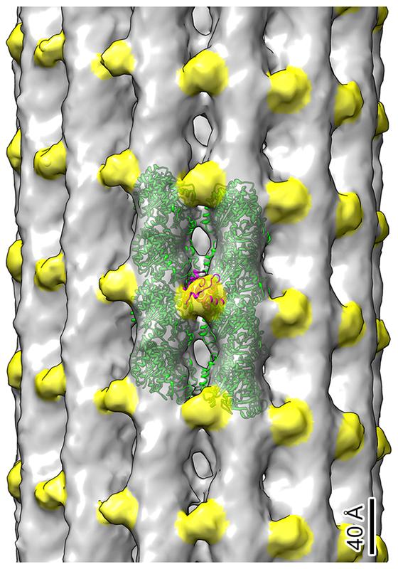
Cryo-electron tomography showing tubulin dimers (in green) and EB1 proteins (in yellow) located along the microtubules of the kidney primary cilium. The functional role of this peculiar distribution of EB1 is now under investigation.
Kiesel et al. in Nature Structural & Molecular Biology / MPI-CBG
Dresden researchers develop a new method and discover new features of primary cilia – little understood antenna-like structures protruding from cells.
Cells sense their environment and send signals to other cells to function properly. The responsible “organ” to perform these functions is the cilium, an antenna-like structure protruding from most vertebrate cells. There are two types of cilia: motile and non-motile cilia. While motile cilia have been studied extensively, the three-dimensional architecture and molecular composition of non-motile cilia, also called primary cilia, are largely unexplored.
Virtually all mammalian cell types have a primary cilium. The research group of Gaia Pigino at the Max Planck Institute of Molecular Cell Biology and Genetics (MPI-CBG) in Dresden has now developed a method for investigating the structure of primary cilia at molecular resolution by cryo-electron tomography. This work provides important insides into the biology of primary cilia and a methodological framework to further study these organelles in various healthy and pathological contexts.
When we explore our environment, we rely on senses like hearing, seeing, tasting, or smelling. A cell senses their surroundings through cilia, antenna-like structures that protrude from most vertebrate cells. Cilia enable cells to move, to communicate, and to interpret molecular signals. Inside a cilium is a microtubule-based cytoskeleton that is studied fairly well in motile cilia.
The same is not true for primary cilia, which have long been seen as a remnant of evolution and have falsely been thought of as a simplified version of motile cilia. But unlike motile cilia, which are found on only some cell types in our bodies, most of our cells have a primary cilium. For example, olfactory sensory cilia enable us to smell, photoreceptor cilia sense light and we can see.
Given this crucial role of cilia in our bodies, their malfunctions can cause a wide range of human diseases, including retinal degeneration, polycystic kidney disease, Bardet-Biedl syndrome, or congenital heart disease. It is therefore of critical importance to better understand primary cilia. To date, there is little mechanistic understanding of how primary cilia perform their functions, and what held us back to better understand them are missing methods to inspect their structure at molecular resolution.
With this motivation in mind, research group leader Gaia Pigino and her team at the MPI-CBG set out to explore more about the structure of primary cilia. They started to work on a method that would eventually allow them to see the molecular structure of the primary cilium in high-resolution. The challenge was to separate the cilia from the cells without destroying their many fine structural details, and then look at them under a particularly powerful electron microscope.
Petra Kiesel, the technician of the research group and the person who developed the method, explains: “We combined a variation of an existing chemical method with a mechanical one, in order to isolate the cilia from kidney cells directly on the microscopy support to image them with our favorite electron microscope.”
For this, the researchers used a method called cryo-electron tomography (Cryo-ET), a special technique to image fast-frozen biological samples in a way that full 3D models of seen molecular structure can be created. With this technique, the researchers were able to observe primary cilia at such a high-resolution that individual proteins could be identified.
Gonzalo Alvarez Viar, PhD student in the research group, reports: “The structures we saw are very different from the ones, we know from motile cilia. Their cytoskeleton is less organized and has a less symmetrical structure. Motile cilia are known to have a nine-fold symmetry in order to move around, but we didn’t see anything like this in primary cilia.”
But not only the structure of known components turns out to be different. Primary cilia also surprised with a number of other novelties, for example the presence of actin filaments inside those cilia. Actin Filaments are found in the cytoplasm of eukaryotic cells and form part of the cytoskeleton. “Finding filamentous actin in cilia was a huge surprise to us. It was sometimes hinted at by other experiments, but only few people really believed in it since there has never been solid evidence”, says Gaia Pigino who oversaw the study.
All findings, revealing a lot of previously unknown facts and surprising differences between the well-studied motile cilia and little understood primary cilia, are now published in Nature Structural and Molecular Biology. “Our method will pave the way for many insightful investigations of primary cilia and, in turn, allow us to better understand these important organelles in animals and humans, in health and disease”, concludes Gaia Pigino.
Wissenschaftliche Ansprechpartner:
Dr. Gaia Pigino
+49 (0) 351 210 2450
pigino@mpi-cbg.de
Originalpublikation:
Petra Kiesel, Gonzalo Alvarez Viar, Nikolai Tsoy, Riccardo Maraspini, Peter Gorilak, Vladimir Varga, Alf Honigmann, Gaia Pigino: “The molecular structure of mammalian primary cilia revealed by cryo-electron tomography”, Nat Struct Mol Biol, 28. September 2020. Doi: 10.1038/s41594-020-0507-4
Media Contact
All latest news from the category: Life Sciences and Chemistry
Articles and reports from the Life Sciences and chemistry area deal with applied and basic research into modern biology, chemistry and human medicine.
Valuable information can be found on a range of life sciences fields including bacteriology, biochemistry, bionics, bioinformatics, biophysics, biotechnology, genetics, geobotany, human biology, marine biology, microbiology, molecular biology, cellular biology, zoology, bioinorganic chemistry, microchemistry and environmental chemistry.


Newest articles
Red light therapy for repairing spinal cord injury passes milestone
Patients with spinal cord injury (SCI) could benefit from a future treatment to repair nerve connections using red and near-infrared light. The method, invented by scientists at the University of…
Insect research is revolutionized by technology
New technologies can revolutionise insect research and environmental monitoring. By using DNA, images, sounds and flight patterns analysed by AI, it’s possible to gain new insights into the world of…

X-ray satellite XMM-newton sees ‘space clover’ in a new light
Astronomers have discovered enormous circular radio features of unknown origin around some galaxies. Now, new observations of one dubbed the Cloverleaf suggest it was created by clashing groups of galaxies….

