A neuromuscular model for drug development

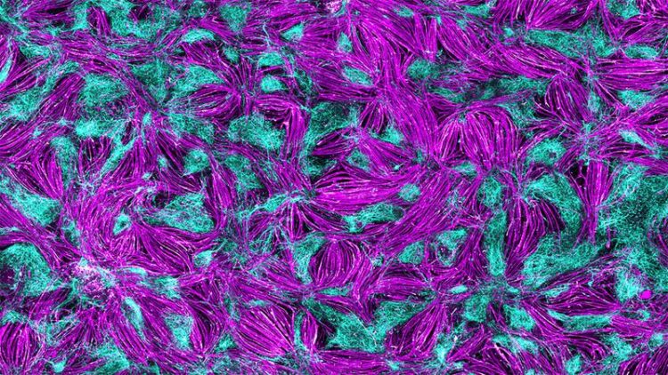
A human self-organizing 2D neuromuscular junction model. Immunofluorescence analysis of the whole dish shows muscle cells (magenta) organized in bundles surrounded by spinal cord neurons (cyan).
Credit: Alessia Urzi, Max Delbrück Center
Scientists have so far identified around 800 different neuromuscular diseases. These conditions are caused by problems in the way muscle cells, motor neurons and peripheral cells interact. These disorders, including amyotrophic lateral sclerosis and spinal muscular atrophy, lead to muscle weakness, paralysis, and in some cases death.
“These diseases are highly complex, and the causes of the dysfunction can vary widely,” says Dr. Mina Gouti, head of the Stem Cell Modeling of Development and Disease Lab at the Max Delbrück Center. The problem might lie with the neurons, the muscle cells or the connections between the two. “To better understand the causes and find effective therapies, we need human-specific cell culture models where we can study how motor neurons in the spinal cord interact with the muscle cells.”
Organoids are too large for high-throughput studies
The researchers working with Gouti had already developed a three-dimensional neuromuscular organoid (NMO) system. “One of our goals is to use our cultures for large-scale drug testing,” says Gouti. “The three-dimensional organoids are very large and can’t be grown for a long time in the 96 well culture dish that we use to perform high-throughput drug screening studies.”
For this type of screening, an international team led by Gouti has now developed a self-organizing neuromuscular junction model using pluripotent stem cells. The model contains neurons, muscle cells, and the chemical synapse named neuromuscular junction that is needed for the two types of cells to interact. The researchers have now published their findings in Nature Communications. “The 2D self-organizing neuromuscular junction model will allow us to perform high throughput drug screening for different neuromuscular diseases and then study the most promising candidates in patient-specific organoids,” says Gouti.
To establish the 2D self-organizing neuromuscular junction model, the researchers first had to understand how motor neurons and muscle cells develop in the embryo. Minas’ team does not conduct embryonic research themselves but utilizes various human stem cell lines, which are allowed for research purposes under strict guidelines, as well as an induced pluripotent stem cell line (iPSC). “We tested a number of hypotheses. We found that the types of cells we needed for functional neuromuscular connections originated from neuromesodermal progenitor cells,” says Alessia Urzi, a doctoral student and lead author of the paper. Urzi found the right combination of signaling molecules that cause human stem cells to mature into functional motor neurons and muscle cells with the necessary connections between the two. “It was exciting to see the muscle cells contracting under the microscope,” says Urzi. “That was a clear sign we were on the right track.” Another observation was that, once differentiated, the cells organized themselves into areas with muscle cells and nerve cells, rather like a mosaic.
An optogenetic switch for motor neurons
The muscle cells grown in the culture dish contract spontaneously as a result of their connection to the neurons – but they do so without any meaningful rhythm. Urzi and Gouti wanted to fix that. Working with researchers at Charité – Universitätsmedizin Berlin, they used optogenetics to activate the motor neurons. Activated by a flash of light, the neurons fire and cause the muscle cells to contract in sync, moving them closer to mimicking the physiological situation in an organism.
Modeling Spinal muscular atrophy in the dish
To test the validity of the model, Urzi used human iPSCs from patients with Spinal muscular atrophy, a severe neuromuscular disease that affects children in the first year of their life. The neuromuscular cultures generated from the patient-specific induced pluripotent stem cells showed severe problems with the contraction of the muscle resembling the patient’s pathology.
For Gouti, the 2D and 3D cultures are key tools for researching neuromuscular diseases in greater detail and to test more efficient and individualized treatment options. As a next step, Gouti and her team want to perform high throughput drug screening to identify novel treatments for patients with spinal muscular atrophy and amyotrophic lateral sclerosis. “We want to start by seeing if we can achieve more successful outcomes using new combinations of drugs to improve the life of patients with complex neuromuscular diseases,” says Gouti.
Further information
Neuromuscular organoid: It’s contracting!
Mina Gouti’s lab
Literature
Alessia Urzi et al. (2023): “Efficient generation of a self-organizing neuromuscular junction model from human pluripotent stem cells”, Nature Communications, DOI: 10.1038/s41467-023-43781-3
Media to download
Picture: A human self-organizing 2D neuromuscular junction model. Immunofluorescence analysis of the whole dish shows muscle cells (magenta) organized in bundles surrounded by spinal cord neurons (cyan). Credit: Alessia Urzi, Max Delbrück Center
Video: A human self-organizing 2D neuromuscular junction model from human pluripotent stem cells. Credit: Gouti Lab, Max Delbrück Center
Contacts
Dr. Mina Gouti
Head of the Stem Cell Modeling of Development and Disease Lab
Max Delbrück Center
+49 (0)30 9406-2610
Christina Anders
Editor, Communications Department
Max Delbrück Center
+49-(0)30-9406-2118
christina.anders@mdc-berlin.de or presse@mdc-berlin.de
Max Delbrück Center
The Max Delbrück Center for Molecular Medicine in the Helmholtz Association (Max Delbrück Center) is one of the world’s leading biomedical research institutions. Max Delbrück, a Berlin native, was a Nobel laureate and one of the founders of molecular biology. At the locations in Berlin-Buch and Mitte, researchers from some 70 countries study human biology – investigating the foundations of life from its most elementary building blocks to systems-wide mechanisms. By understanding what regulates or disrupts the dynamic equilibrium of a cell, an organ, or the entire body, we can prevent diseases, diagnose them earlier, and stop their progression with tailored therapies. Patients should be able to benefit as soon as possible from basic research discoveries. This is why the Max Delbrück Center supports spin-off creation and participates in collaborative networks. It works in close partnership with Charité – Universitätsmedizin Berlin in the jointly-run Experimental and Clinical Research Center (ECRC), the Berlin Institute of Health (BIH) at Charité, and the German Center for Cardiovascular Research (DZHK). Founded in 1992, the Max Delbrück Center today employs 1,800 people and is 90 percent funded by the German federal government and 10 percent by the State of Berlin.
Journal: Nature Communications
DOI: 10.1038/s41467-023-43781-3
Article Title: Efficient generation of a self-organizing neuromuscular junction model from human pluripotent stem cells
Article Publication Date: 19-Dec-2023
Media Contact
All latest news from the category: Life Sciences and Chemistry
Articles and reports from the Life Sciences and chemistry area deal with applied and basic research into modern biology, chemistry and human medicine.
Valuable information can be found on a range of life sciences fields including bacteriology, biochemistry, bionics, bioinformatics, biophysics, biotechnology, genetics, geobotany, human biology, marine biology, microbiology, molecular biology, cellular biology, zoology, bioinorganic chemistry, microchemistry and environmental chemistry.
Newest articles

Recovering phosphorus from sewage sludge ash
Chemical and heat treatment of sewage sludge can recover phosphorus in a process that could help address the problem of diminishing supplies of phosphorus ores. Valuable supplies of phosphorus could…

Efficient, sustainable and cost-effective hybrid energy storage system for modern power grids
EU project HyFlow: Over three years of research, the consortium of the EU project HyFlow has successfully developed a highly efficient, sustainable, and cost-effective hybrid energy storage system (HESS) that…

After 25 years, researchers uncover genetic cause of rare neurological disease
Some families call it a trial of faith. Others just call it a curse. The progressive neurological disease known as spinocerebellar ataxia 4 (SCA4) is a rare condition, but its…


