High-resolution 3D view inside breast tumors with opto-acoustic mesoscopy
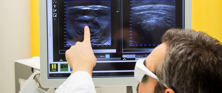
Prof. Vasilis Ntziachristos demonstrates an imaging method similar to multi-spectral optoacoustic mesoscopy. Image: M. Jooss
Malignant tumors consume nutrients and oxygen faster than healthy cells. To do so, they recruit blood vessels in their environment. Depending on tumor type and genetic profile, there are differences on how tumors look internally.
Typically, tumors present different patterns across their volume. The role of this spatial heterogeneity is not well understood or studied in living tumors.
Typically used to understand biological functions in tumors, optical microscopy, for example, gives limited insights into the spatial heterogeneity of tumors as it only accesses volumes of less than a cubic millimeter.
High resolution with new imaging method
A new technique developed by Munich researchers, known as multi-spectral optoacoustic mesoscopy (MSOM), has now been shown capable of resolving optical contrast through tumor volumes that are at least 1,000 times larger than those possible with optical microscopy, enabling high-resolution visualization of tumor heterogeneity patterns.
With this imaging method, the tumor is first excited from all sides with pulses of infrared laser light. “Tumor and tissue components that absorb this excitation light undergo a tiny, transient temperature increase, which leads to a small local volume expansion, followed by a contraction. This expansion and contraction process generates a weak ultrasound signal, which we collect with a detector,” says guest researcher Dr. Jiao Li.
The data collected is mathematically processed to form light absorption images which indicate different tumor patterns reflecting tumor oxygenation and vascularization. “For the first time, MSOM offers optical images that reach inside tumors to depths of ten millimeters and more with a resolution of less than 50 micrometers,” says Dr. Li.
Understanding functional variety in tumors
“MSOM imaging of solid tumors allowed us to see tumors in a new light,” says Prof. Vasilis Ntziachristos, holder of the Chair of Biological Imaging at TUM and Director of the Institute for Biological and Medical Imaging at Helmholtz Zentrum München. “MSOM allows us to understand how tumor functionality varies across the tumor, clearly moving the reach of optical observations well beyond the depth penetration limitations of optical microscopy”.
In the pictures taken from mamma carcinomas of mice, researchers can see patterns indicating the presence or absence of blood vessels, and thus study blood supply patterns. MSOM can also resolve hemoglobin levels, and indicate whether oxygen is bound to hemoglobin or not. Furthermore, MSOM images were used to determine permeability of the vessel walls relative to nanoparticles. Using the mouse model, the scientists were already able to track how tiny gold particles were transported.
3D tumor pictures without surgical biopsy
In contrast to conventional histology, where tissue has to be removed, cut up and examined under the microscope by a pathologist, MSOM allows a three-dimensional analysis of entire living tumors without the need for surgical biopsies. This further supports longitudinal studies so that tumor growth or recession under different drugs can be studied with greater accuracy. All of which paves the way for an improved understanding of biological function and drug efficacy during drug development for humans.
More information:
The research work was supported by the German Research Foundation (DFG), the European Research Council (ERC) and the National Natural Science Foundation of China.
Prof. Dr. Vasilis Ntziachristos
Technical University of Munich
Chair of Biological Imaging
Phone: +49 89 4140 7211
v.ntziachristos@tum.de
J. Li, A. Chekkoury, J. Prakash, S. Glasl, P. Vetschera, B. Koberstein-Schwarz, I. Olefir, V. Gujrati, M. Omar, V. Ntziachristos: Spatial heterogeneity of oxygenation and hemodynamics in breast cancer resolved in vivo by conical multispectral optoacoustic mesoscopy. Light Sci Appl 9, 57 (2020). DOI: 10.1038/s41377-020-0295-y
Media Contact
More Information:
https://www.tum.de/nc/en/about-tum/news/press-releases/details/36044/All latest news from the category: Medical Engineering
The development of medical equipment, products and technical procedures is characterized by high research and development costs in a variety of fields related to the study of human medicine.
innovations-report provides informative and stimulating reports and articles on topics ranging from imaging processes, cell and tissue techniques, optical techniques, implants, orthopedic aids, clinical and medical office equipment, dialysis systems and x-ray/radiation monitoring devices to endoscopy, ultrasound, surgical techniques, and dental materials.


Newest articles

A universal framework for spatial biology
SpatialData is a freely accessible tool to unify and integrate data from different omics technologies accounting for spatial information, which can provide holistic insights into health and disease. Biological processes…

How complex biological processes arise
A $20 million grant from the U.S. National Science Foundation (NSF) will support the establishment and operation of the National Synthesis Center for Emergence in the Molecular and Cellular Sciences (NCEMS) at…

Airborne single-photon lidar system achieves high-resolution 3D imaging
Compact, low-power system opens doors for photon-efficient drone and satellite-based environmental monitoring and mapping. Researchers have developed a compact and lightweight single-photon airborne lidar system that can acquire high-resolution 3D…

