Proteins imaged in graphene liquid cell have higher radiation tolerance
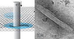
Schematic representation
Electron microscopy is one of the main methods used to examine protein structure. Studying these structures is of key importance to elucidate their function feeding fundamental information into a number of fields such as structural biology, cell biology, cancer research, and other biomedical fields. It also enhances the understanding of biomineralization.
A new option for imaging proteins is liquid-phase electron microscopy (LPEM), which is capable of imaging native (unstained) protein structure and other samples such as nanomaterials or cells in liquid.
This technology was developed over the past fifteen years. Until recently, it debated whether the radiation tolerance of liquid samples would be better or worse compared to amorphous ice.
In their recent publication, Sercan Keskin and Niels de Jonge from the INM-Leibniz Institute for New Materials now demonstrate, that the radiation tolerance is increased by an order of magnitude compared to a sample in ice.
This result was achieved by preparing a microtubule sample in a graphene liquid cell. Essential was to use a low as possible rate at which the electron beam irradiation was applied.
Traditionally, samples were fixed, stained with a metal to enhance their contrast, subsequently dried, embedded in plastic, cut in thin sections, and then imaged in the vacuum environment required for electron microscopy.
Cryo electron microscopy overcomes the drawbacks associated with this sample preparation and provides the means to study proteins in a close to native hydrated state by preparing them in amorphous ice.
However, a key imitating is the high sensitivity of the samples to electron beam irradiation, so that statistical noise in the image prevents high resolution and many ten thousand noisy images of identical structures need to be imaged in order to resolve the structure.
Prof. Dr. Niels de Jonge
Head Innovative Electron Microscopy
Phone: +49681-9300-313
niels.dejonge@leibniz-inm.de
Keskin, S. & de Jonge, N. Reduced radiation damage in transmission electron microscopy of proteins in graphene liquid cells. Nano Lett., early online, 2018. DOI: 10.1021/acs.nanolett.8b02490
Media Contact
More Information:
http://www.inm-gmbh.deAll latest news from the category: Materials Sciences
Materials management deals with the research, development, manufacturing and processing of raw and industrial materials. Key aspects here are biological and medical issues, which play an increasingly important role in this field.
innovations-report offers in-depth articles related to the development and application of materials and the structure and properties of new materials.


Newest articles

Sea slugs inspire highly stretchable biomedical sensor
USC Viterbi School of Engineering researcher Hangbo Zhao presents findings on highly stretchable and customizable microneedles for application in fields including neuroscience, tissue engineering, and wearable bioelectronics. The revolution in…

Twisting and binding matter waves with photons in a cavity
Precisely measuring the energy states of individual atoms has been a historical challenge for physicists due to atomic recoil. When an atom interacts with a photon, the atom “recoils” in…
Nanotubes, nanoparticles, and antibodies detect tiny amounts of fentanyl
New sensor is six orders of magnitude more sensitive than the next best thing. A research team at Pitt led by Alexander Star, a chemistry professor in the Kenneth P. Dietrich…

