Single-neuron observations mark steps in Alzheimer's disease
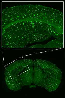
These are microscopic images of a brain section (bottom: overview. top: enlarged view of the visual cortex), obtained from an APP23xPS45 Alzheimer mouse, in which the beta-amyloid plaques are labeled with Thioflavin-S. Credit: Konnerth lab, TU Muenchen<br>
Studying a mouse model of Alzheimer's disease, neuroscientists at the Technische Universitaet Muenchen have observed correlations between increases in both soluble and plaque-forming beta-amyloid – a protein implicated in the disease process – and dysfunctional developments on several levels: individual cortical neurons, neuronal circuits, sensory cognition, and behavior.
Their results, published in Nature Communications, show that these changes progress in parallel and that, together, they reveal distinct stages in Alzheimer's disease with a specific order in time.
In addition to its well known, devastating effects on memory and learning, Alzheimer's disease can also impair a person's sense of smell or vision. Typically these changes in sensory cognition only show themselves behaviorally when the disease is more advanced. A new study sheds light on what is happening in the brain throughout the disease process, specifically with respect to the part of the cerebral cortex responsible for integrating visual information. A team led by Prof. Arthur Konnerth, a Carl von Linde Senior Fellow of the TUM Institute for Advanced Study, has observed Alzheimer's-related changes in the visual cortex at the single-cell level.
Using a technique called two-photon calcium imaging, the researchers recorded both spontaneous and stimulated signaling activity in cortical neurons of living mice: transgenic mice carrying mutations that cause Alzheimer's disease in humans, and wild-type mice as a control group. By observing how neuronal signaling responded to a special kind of vision test – in which a simple grating pattern of light and dark bars moves in front of the mouse's eye – the scientists could characterize the visual circuit as being more or less “tuned” to specific orientations and directions of movement.
Konnerth explains, “Like many Alzheimer's patients, the diseased mice have impairments in their ability to discriminate visual objects. Our results provide important new insights on the cause that may underlie the impaired behavior, by identifying in the visual cortex a fraction of neurons with a strongly disturbed function.” And within this group, the researchers discovered, there are two subsets of neurons – both dysfunctional, but in completely different ways. One subset, thought to be the first neurons to degenerate, showed no activity at all; the other showed a pathologically high level of activity, rendering these neurons incapable of properly sensing objects in the mouse's environment. “While around half of the neurons in the visual cortex were disturbed in one way or the other, roughly half responded normally,” notes Christine Grienberger, a doctoral candidate in Konnerth's institute and first author of this paper. “That could have significant implications for future research in the field of Alzheimer's disease, as our findings raise the question of whether future work only needs to target this population of neurons that are disturbed in their function.”
The in vivo single-neuron experiments were carried out for three age groups, corresponding to different stages of this progressive, degenerative disease. The results were correlated with other measurements, including soluble beta-amyloid levels and the density of beta-amyloid plaques in the brain tissue. The researchers' findings show for the first time a progressive decline of function in cortical circuits. “An important conclusion from this study,” Konnerth says, “is that the Alzheimer's disease-related changes on all levels – including behavior, cortical circuit dysfunction, and the density of amyloid plaques in diseased brains – progress in parallel in a distinct temporal order. In the future, the identification of such stages in patients may help researchers pinpoint stage-specific and effective therapies, with reduced levels of side effects.”
This research was supported by the German Excellence Initiative (TUM-IAS, CIPSM); the German Research Foundation (DFG, IRTG 1373); ERAnet; and the Friedrich Schiedel Foundation.
Original publication:
Staged decline of neuronal function in vivo in an animal model of Alzheimer's disease
Christine Grienberger, Nathalie L. Rochefort, Helmuth Adelsberger, Horst A. Henning, Daniel N. Hill, Julia Reichwald, Matthias Staufenbiel, and Arthur Konnerth
Nature Communications, April 10, 2012.
DOI: 10.1038/ncomms1783
Contact:
Prof. Arthur Konnerth
Institute for Neuroscience
Technische Universitaet Muenchen
Biedersteinerstr. 29, 80802 Munich, Germany
Tel.: +49 (0)89 4140 3351
E-Mail: arthur.konnerth@lrz.tum.de
Web: http://www.ifn.me.tum.de/new/konnerth.html
Technische Universitaet Muenchen (TUM) is one of Europe's leading universities. It has roughly 460 professors, 9000 academic and non-academic staff, and 31,000 students. It focuses on the engineering sciences, natural sciences, life sciences, medicine, and economic sciences. After winning numerous awards, it was selected as an “Elite University” in 2006 by the Science Council (Wissenschaftsrat) and the German Research Foundation (DFG). The university's global network includes an outpost with a research campus in Singapore. TUM is dedicated to the ideal of a top-level research-based entrepreneurial university.
Media Contact
More Information:
http://www.tum.deAll latest news from the category: Health and Medicine
This subject area encompasses research and studies in the field of human medicine.
Among the wide-ranging list of topics covered here are anesthesiology, anatomy, surgery, human genetics, hygiene and environmental medicine, internal medicine, neurology, pharmacology, physiology, urology and dental medicine.


Newest articles

Sea slugs inspire highly stretchable biomedical sensor
USC Viterbi School of Engineering researcher Hangbo Zhao presents findings on highly stretchable and customizable microneedles for application in fields including neuroscience, tissue engineering, and wearable bioelectronics. The revolution in…

Twisting and binding matter waves with photons in a cavity
Precisely measuring the energy states of individual atoms has been a historical challenge for physicists due to atomic recoil. When an atom interacts with a photon, the atom “recoils” in…
Nanotubes, nanoparticles, and antibodies detect tiny amounts of fentanyl
New sensor is six orders of magnitude more sensitive than the next best thing. A research team at Pitt led by Alexander Star, a chemistry professor in the Kenneth P. Dietrich…

