Observing brain network dynamics to diagnose Alzheimer's disease
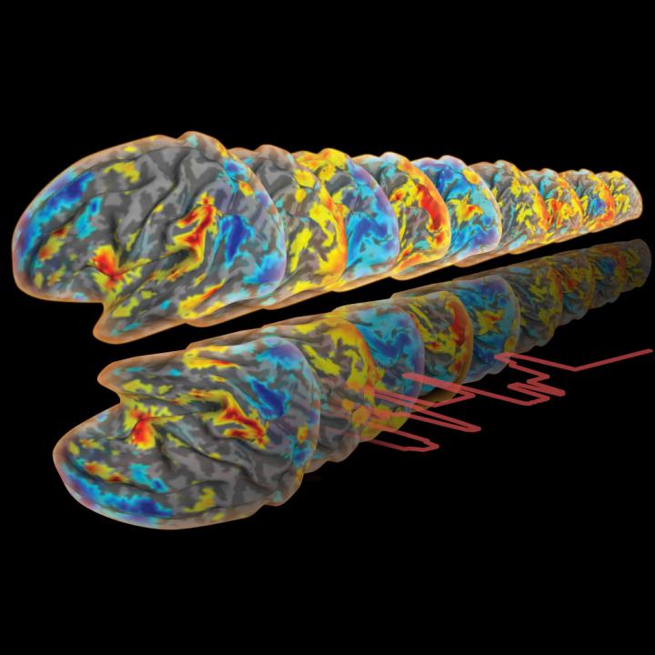
The new imaging technique could help with the early detection of Alzheimer's disease. Credit: EPFL/Dimitri Van De Ville
On the basis of this principle, researchers Isik Karahanoglu and Dimitri Van De Ville have managed to visualize the different activation regions of the brain. They combined a new modeling technique and a medical imaging technique in a project bridging EPFL and the University of Geneva (UNIGE).
The research, published in Nature Communications, provides new insights into how the brain organizes itself, and sets the stage for early diagnosis of neurological disorders like Alzheimer's, in which these networks break down.
In most brain-related disorders, several neural networks – rather than an isolated region – break down. Understanding how the regions interact provides insight into how these disorders work.
Seeing if a region is in “on” or “off” mode
There is already an imaging technique called “functional magnetic resonance imaging” (fMRI), which records variations in blood flow. But this process has its flaws. Thanks to a complex computational method, the researchers were able to clean up the imperfect signals obtained from fMRI and get a precise and dynamic picture of blood flow in the brain. They can see which regions of the brain are activated in an explicit “on” or “off” mode.
“Imagine taking pictures of a rainbow-coloured windmill that is turning very fast. With the old technique, the colours are fuzzy and run together,” said Van De Ville. “With our method we can clearly see the border between each colour on each photo.” Similarly, the dynamic map shows which regions activate simultaneously in the brain and where they are located.
Non-stimulated brain for better data gathering
To identify the regions that work together, the tests were done on healthy, non-stimulated subjects. Even when in a state of 'rest' and not being used, a patient's brain has regions that are constantly activating and deactivating. “The patient must not do anything once in the MRI machine. The data are thus not distorted by the stress or fatigue that stimulation or a task could cause,” said Karahanoglu.
Surprising results
In all, the researchers identified 13 main networks, i.e. those that send out the strongest signals. On average, four of these networks were active at the same time. “Until now, we thought the regions took turns activating, and that they did so with little coordination,” added Van De Ville.
A diagnostic tool for doctors
The next step consists in using this technique to diagnose neurological disorders. Alzheimer disease, for example shows deterioration in brain networks even when clinical symptoms are undetectable or negligible. Using fMRI to detect, as early as possible, cases that are most likely to develop into Alzheimer's would improve drug administration. Drugs currently in development could then be administered during the phase in which they would be most effective. Research along these lines is underway in collaboration with other neuroscience and clinical teams. Isik Karahanoglu, who is currently a post-doctoral fellow at Harvard Medical School, is also applying the technique to better understand alterations in Autism Spectrum Disorder.
This research was made possible through support from the Swiss National Science Foundation, the Bertarelli Foundation and the Center for Biomedical Imaging (CIBM).
Media Contact
All latest news from the category: Health and Medicine
This subject area encompasses research and studies in the field of human medicine.
Among the wide-ranging list of topics covered here are anesthesiology, anatomy, surgery, human genetics, hygiene and environmental medicine, internal medicine, neurology, pharmacology, physiology, urology and dental medicine.


Newest articles

Webb captures top of iconic horsehead nebula in unprecedented detail
NASA’s James Webb Space Telescope has captured the sharpest infrared images to date of a zoomed-in portion of one of the most distinctive objects in our skies, the Horsehead Nebula….
Cost-effective, high-capacity, and cyclable lithium-ion battery cathodes
Charge-recharge cycling of lithium-superrich iron oxide, a cost-effective and high-capacity cathode for new-generation lithium-ion batteries, can be greatly improved by doping with readily available mineral elements. The energy capacity and…
Novel genetic plant regeneration approach
…without the application of phytohormones. Researchers develop a novel plant regeneration approach by modulating the expression of genes that control plant cell differentiation. For ages now, plants have been the…

