


Feeding 9 Billion: Innovations in Agricultural Modeling
LMU researchers have developed a method to determine how reliably target proteins can be labeled using super-resolution fluorescence microscopy.
Modern microscopy techniques make it possible to examine the inner workings of cells in astonishing detail. “We can now observe the arrangement and interaction of individual proteins under the microscope,” says Professor Ralf Jungmann, Chair of Molecular Physics of Life at LMU and Max Planck Fellow at the MPI of Biochemistry. The biophysicist’s team recently developed the revolutionary RESI (Resolution Enhancement by Sequential Imaging) method. This technique can be used to improve the resolution of fluorescence microscopy down to the Ångström scale – far below the classical diffraction limit of light. DNA-conjugated marker molecules, which the researchers attach precisely to the molecules they want to understand better, are crucial for this.
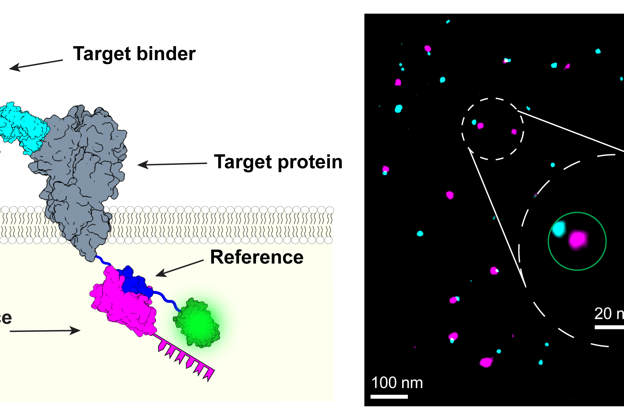
Jungmann’s team has now presented a technique in the journal Nature Methods that can be used to quantify how well biomarker molecules bind to the target proteins. “This is absolutely crucial if you want to make quantitatively reliable statements,” explains the physicist. If you know the labeling efficiency, you can carry out spatially resolved proteomics in this way. This allows you to find out not only what individual proteins do in a cell, but also to what extent they are present and how their quantity and behavior change under certain circumstances. “But this is only possible if we can assess how well the labeling has worked.” This is because only labeled proteins emit flashes of light under the microscope and thus become visible.
The method developed by Jungmann’s team makes this assessment possible by adding a reference biomarker to the target proteins. This marker “glows” in a different color during microscopy, so that successfully marked proteins appear in two colors. Jungmann’s team demonstrated this using the membrane protein CD86, among others: The reference produces a pink fluorescence, the actual marker a bluish one. This creates a pattern of innumerable pink and blue points of light. Where the marking did not work, only the reference lights up individually. The marking efficiency is calculated from the ratio of double and single illuminated molecules.
The technique can be applied to a variety of different target molecules, biomarkers, and samples and is compatible with a whole range of super-resolution microscopy methods. Ralf Jungmann
The authors of the study are certain that the new quantification method has paved the way for significantly expanding the potential of their super-resolution microscope method: “Now we can also consider specific biomedical applications in which the quantitative detection of proteins and processes is of great importance,” says Jungmann. This includes cancer research, for example, where information about interactions between proteins on the cell surface and drugs with molecular resolution is essential for the development of new types of medication.
Joschka Hellmeier, Sebastian Strauss, Shuhan Xu, Luciano A. Masullo, Eduard M. Unterauer, Rafal Kowalewski & Ralf Jungmann: Quantification of absolute labeling efficiency at the single-protein level. Nature Methods, 2024.
https://www.lmu.de/en/newsroom/news-overview/news/biophysics-testing-how-well-biomarkers-work.html


The appearance of a hot sauce or pepper doesn’t reveal whether it’s mild or likely to scorch someone’s taste buds. So, researchers made an artificial tongue to quickly detect spiciness. Inspired by milk’s casein proteins, which bind to capsaicin and relieve the burn of spicy foods, the researchers incorporated milk powder into a gel sensor. The prototype, reported in ACS Sensors, detected capsaicin and pungent-flavored compounds (like those behind garlic’s zing) in various foods. “Our flexible artificial tongue holds tremendous…
Unless you’ve owned reptiles, you might not know that many of them “pee” crystals. Researchers publishing in the Journal of the American Chemical Society investigated the solid urine of more than 20 reptile species and found spheres of uric acid in all of them. This work reveals how reptiles uniquely package up and eliminate crystalline waste, which could inform future treatments for human conditions that also involve uric acid crystals: kidney stones and gout. Most living things have some sort…
Enabled by a new high-resolution mapping technique, the findings overturn a long-held belief that the genome loses its 3D structure when cells divide CAMBRIDGE, MA — Before cells can divide, they first need to replicate all of their chromosomes, so that each of the daughter cells can receive a full set of genetic material. Until now, scientists had believed that as division occurs, the genome loses the distinctive 3D internal structure that it typically forms. Once division is complete, it…
Researchers at Chalmers University of Technology in Sweden and the US space agency NASA have made an unexpected discovery that challenges one of the basic rules of chemistry and provides new knowledge about Saturn’s enigmatic moon Titan. In its extremely cold environment, normally incompatible substances can still be mixed. This discovery broadens our understanding of chemistry before the emergence of life. Scientists have long been interested in Saturn’s largest, orange-coloured moon as its evolution can teach us more about our…
