Technique used commonly in physics finds application in neuroscience
To understand how brain cells release compounds (or transmitters) used when the cells communicate with each other, Vladimir Parpura, associate professor of neuroscience, and Umar Mohideen, professor of physics at UC Riverside, devised a new technique, used commonly in physics, that can be applied now to the study of a wide range of biological processes and interactions.
The researchers, who performed their experiments on brain proteins called SNAREs, published their results in the July issue of Biophysical Journal.
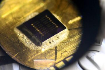
The technique, commonly referred to as Atomic Force Microscopy, uses the deflection of microfabricated membranes of silicon nitride, about 100 times thinner than the human hair, to measure very small forces. Using this technique on rat brain proteins, the researchers were able to measure the bonding between single protein molecules that are involved in the release of the neurotransmitters. They also were able to classify the strength of the molecular interactions (bonding) between 3 of the SNARE proteins that participate in the process.
SNARE proteins are located on vesicles (tiny membrane-encased packets that contain neurotransmitters or enzymes) and the plasma membrane of brain cells. These proteins are thought to play a key role in the final fusion of the synaptic vesicle with the plasma membrane, a process that makes communication between cells possible.
“Our results shed new light on how these proteins are involved in exocytosis – the process by which a biological cell releases substances into its environment,” Parpura said. “We now understand better how these proteins interact at the molecular level and we can apply this to improve our detection of toxins acting on these proteins.”
The researchers used the technique also to develop a sensor for detecting botulinum toxin, responsible for an often fatal type of food poisoning.
“Our sensor is extremely sensitive because it is capable of detecting interactions between two single molecules,” Mohideen said. “As a result, the sample size you need for testing can be extremely small, of the order of a few molecules.”
Media Contact
More Information:
http://www.ucr.eduAll latest news from the category: Medical Engineering
The development of medical equipment, products and technical procedures is characterized by high research and development costs in a variety of fields related to the study of human medicine.
innovations-report provides informative and stimulating reports and articles on topics ranging from imaging processes, cell and tissue techniques, optical techniques, implants, orthopedic aids, clinical and medical office equipment, dialysis systems and x-ray/radiation monitoring devices to endoscopy, ultrasound, surgical techniques, and dental materials.


Newest articles

Sea slugs inspire highly stretchable biomedical sensor
USC Viterbi School of Engineering researcher Hangbo Zhao presents findings on highly stretchable and customizable microneedles for application in fields including neuroscience, tissue engineering, and wearable bioelectronics. The revolution in…

Twisting and binding matter waves with photons in a cavity
Precisely measuring the energy states of individual atoms has been a historical challenge for physicists due to atomic recoil. When an atom interacts with a photon, the atom “recoils” in…
Nanotubes, nanoparticles, and antibodies detect tiny amounts of fentanyl
New sensor is six orders of magnitude more sensitive than the next best thing. A research team at Pitt led by Alexander Star, a chemistry professor in the Kenneth P. Dietrich…

