New Computer Technique Differentiates Malignant and Benign Calcifications on Digital Mammograms

Researchers at the University of Chicago have developed a computer technique that “learns” how benign and malignant breast calcifications appear on digital mammograms so not only can it detect them, but it can also predict the likelihood that the calcifications are associated with cancer.
“In this study, we analyzed 49 full-field digital mammograms, 19 of which showed cancer,” said Rich Rana, a medical student at the University of Chicago. Four mammography specialists read the images and electronically put a box around the suspicious calcifications. The computer then automatically detected the calcifications within the box, analyzed them and calculated the probability of cancer, Rana said.
The system proved to consistently achieve performance comparable to the radiologists in classifying malignant and benign calcifications, regardless of who was using it, Rana added. One technique for rating the computer’s effectiveness is to give it one malignant case and one benign case and then test its ability to determine which is which, Rana said. Using this technique, the radiologist had a 72% chance of making the correct diagnosis, and the computer had a 79% chance.
This study was one of the first to test the effectiveness of computer-aided diagnoses on full-field digital mammograms versus plain film mammograms. In addition, this system employs artificial intelligence in that the computer “learns” how to automatically locate the calcifications and predict whether they are benign or malignant, Rana said. In the future the radiologist’s assessment could be compared with the computer’s assessment as a “double-check” for the diagnosis of breast cancer, Rana said.
This research was lead by Dr. Yulei Jiang, assistant professor of radiology at the University of Chicago. The research was funded by the National Cancer Institute, the U.S. Army Medical Research and Material Command, and the National Institutes of Health. Rana will present the study on May 4 at the American Roentgen Ray Society Annual Meeting in Miami Beach, FL.
Media Contact
More Information:
http://www.arrs.org/scriptcontent/pressroom/archive/2004/r040504g.cfmAll latest news from the category: Health and Medicine
This subject area encompasses research and studies in the field of human medicine.
Among the wide-ranging list of topics covered here are anesthesiology, anatomy, surgery, human genetics, hygiene and environmental medicine, internal medicine, neurology, pharmacology, physiology, urology and dental medicine.
Newest articles

A universal framework for spatial biology
SpatialData is a freely accessible tool to unify and integrate data from different omics technologies accounting for spatial information, which can provide holistic insights into health and disease. Biological processes…

How complex biological processes arise
A $20 million grant from the U.S. National Science Foundation (NSF) will support the establishment and operation of the National Synthesis Center for Emergence in the Molecular and Cellular Sciences (NCEMS) at…

Airborne single-photon lidar system achieves high-resolution 3D imaging
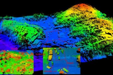
Compact, low-power system opens doors for photon-efficient drone and satellite-based environmental monitoring and mapping. Researchers have developed a compact and lightweight single-photon airborne lidar system that can acquire high-resolution 3D…


