NIST effort could improve high-tech medical scanners
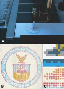
Microarrayer machines (A) now can mix colors and deposit them on microscope slides, which can be used to calibrate hyperspectral imagers (HSI) for use in medical applications. The finished slides can be custom-colored (B) to calibrate HSIs to find specific types of tumors or disease tissue. Close up, they resemble dot-matrix printwork (C). Credit: Clarke/NIST<br>
The technique, called hyperspectral imaging (HSI), has frequently been used in satellites because of its superior ability to identify objects by color. While many other visual surveying methods can scan only for a single color, HSI is able to distinguish the full color spectrum in each pixel, which allows it to perceive the unique color “signatures” of individual objects.
Well-calibrated HSI sensors have been able to discern problems from diseases in coral reefs to pollution in the atmosphere as determined by the distinct spectral signature at a location.
“Because diseased tissues and cells also have distinct spectra, scientists have been trying to use HSI for medical applications as well,” says NIST physicist Jeeseong Hwang. “But any time you tell a machine to scan for something, you need to be sure it is actually looking for what you want, and you have to make sure that the image analysis algorithm extracts the correct color information out of a complex multicolor data set. We decided to create a way to calibrate an HSI device and to test its algorithm as well.”
Matthew Clarke, a former National Research Council-supported postdoctoral fellow in Hwang's group who is currently working in the National Gallery of Art in Washington, D.C., wrote new software for a device called a microarrayer, so named because it is capable of laying down hundreds of tiny sample droplets in specific places on a microscope slide's surface. Normally a microarrayer creates DNA arrays for genetic research, but the team remade it into an artistic tool, programming it to select chemicals of different hues and lay them down on the slide's surface.
The results, which look a bit like dot-matrix printing, can be used to calibrate medical HSI devices and image analysis algorithms. When combined with HSI in a medical imaging application, this effort could allow a surgeon to look for cells with a specific chemical makeup, as determined by the cells' color.
“Scientists and engineers can create a custom slide with the exact colors representing the chemical makeup they want the HSI devices to detect,” Hwang says. “It could be a good way to make sure the HSI devices for medical imaging perform correctly so that surgeons are able to see all of a tumor or diseased tissue when operating on a patient.”
This project is part of a larger effort to evaluate and validate optical medical imaging devices, led by the NIST team members, David Allen, Maritoni Litorja, Antonio Possolo, Eric Shirley and Jeeseong Hwang. Hwang adds that the special issue** of Biomedical Optics Express in which the team's findings appear is the output of a recent NIST-supported international workshop on the topic.
*M.L. Clarke, J.Y. Lee, D.V. Samarov, D.W. Allen, M. Litorja, R. Nossal and J. Hwang. Designing microarray phantoms for hyperspectral imaging validation. Biomedical Optics Express, Vol. 3(6), pp. 1291-1299 (June 2012), doi: 10.1364/BOE.3.001300.
** See www.opticsinfobase.org/boe/virtual_issue.cfm?vid=168.
Media Contact
More Information:
http://www.nist.govAll latest news from the category: Medical Engineering
The development of medical equipment, products and technical procedures is characterized by high research and development costs in a variety of fields related to the study of human medicine.
innovations-report provides informative and stimulating reports and articles on topics ranging from imaging processes, cell and tissue techniques, optical techniques, implants, orthopedic aids, clinical and medical office equipment, dialysis systems and x-ray/radiation monitoring devices to endoscopy, ultrasound, surgical techniques, and dental materials.


Newest articles

A universal framework for spatial biology
SpatialData is a freely accessible tool to unify and integrate data from different omics technologies accounting for spatial information, which can provide holistic insights into health and disease. Biological processes…

How complex biological processes arise
A $20 million grant from the U.S. National Science Foundation (NSF) will support the establishment and operation of the National Synthesis Center for Emergence in the Molecular and Cellular Sciences (NCEMS) at…

Airborne single-photon lidar system achieves high-resolution 3D imaging
Compact, low-power system opens doors for photon-efficient drone and satellite-based environmental monitoring and mapping. Researchers have developed a compact and lightweight single-photon airborne lidar system that can acquire high-resolution 3D…

