Mapping Atherosclerotic Arteries: Combined Approach Developed
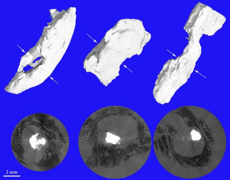
Conventional micro-tomography using intense X-rays allows for the visualization of plaque (white) and muscle tissue (black). Biomaterials Science Center, University of Basel
Cardiovascular diseases, including atherosclerosis, are associated with plaque formation and the most prevalent cause of death worldwide. Unlike vessels and other soft tissues, the plaque formed provides strong contrast in X-rays, as known from bone. So far, it has therefore been difficult or even impossible to identify soft tissues in the direct neighborhood of calcifications using X-rays.
A team of researchers from laboratories in three European countries, led by Bert Müller (Biomaterials Science Center at University of Basel), has developed a protocol that is based on the combination of hard X-ray tomography and established histology methods, to visualize the vessels constricted by atherosclerosis.
The data about the morphology of the constricted vessels is used to simulate blood flow and determine related shear stresses. The shear stress is significantly enhanced at the constrictions and forms the basis for the development of specialized nano-containers for the targeted and local delivery of vasodilation drugs.
Differentiation between soft and hard tissues
The new method combines known approaches and is not only suitable for the three-dimensional characterization of atherosclerotic blood vessels but also for any other combination of strongly and weakly X-ray absorbing species including cartilage and bone. It takes advantage of conventional X-ray absorption and, in addition, of X-ray phase contrast measurements, which are for example accessible via grating interferometry. As the phase contrast is much less dependent on the atomic number of the constituents than the absorption contrast, the soft tissues in the vicinity of hard tissues become much more easily visualized.
In summary, the authors demonstrate that strongly calcified arteries are thoroughly characterized by the combination of the non-destructive tomography measurements in X-ray absorption and phase contrast modes, and established histology techniques. The project “NO-stress” is funded within the National Research Programme NRP 62 “Smart Materials” by the Swiss National Science Foundation.
Original citation
Margaret N Holme, Georg Schulz, Hans Deyhle, Timm Weitkamp, Felix Beckmann, Johannes A Lobrinus, Farhad Rikhtegar, Vartan Kurtcuoglu, Irene Zanette, Till Saxer, Bert Müller
Complementary X-ray tomography techniques for histology-validated three-dimensional imaging of soft and hard human tissues
Nature Protocols 9, 1401-1415 | doi:10.1038/nprot.2014.091
Further information
Prof. Bert Müller, Biomaterials Science Center at the University of Basel, Tel. +41 (0)61 265 96 60, E-Mail: bert.mueller@unibas.ch
http://www.nature.com/nprot/journal/v9/n6/full/nprot.2014.091.html – Full Version
Media Contact
More Information:
http://www.unibas.chAll latest news from the category: Materials Sciences
Materials management deals with the research, development, manufacturing and processing of raw and industrial materials. Key aspects here are biological and medical issues, which play an increasingly important role in this field.
innovations-report offers in-depth articles related to the development and application of materials and the structure and properties of new materials.


Newest articles

A universal framework for spatial biology
SpatialData is a freely accessible tool to unify and integrate data from different omics technologies accounting for spatial information, which can provide holistic insights into health and disease. Biological processes…

How complex biological processes arise
A $20 million grant from the U.S. National Science Foundation (NSF) will support the establishment and operation of the National Synthesis Center for Emergence in the Molecular and Cellular Sciences (NCEMS) at…

Airborne single-photon lidar system achieves high-resolution 3D imaging
Compact, low-power system opens doors for photon-efficient drone and satellite-based environmental monitoring and mapping. Researchers have developed a compact and lightweight single-photon airborne lidar system that can acquire high-resolution 3D…

