Novel technique has potential to transform breast cancer detection
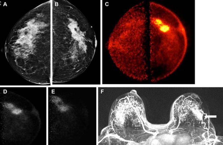
Images obtained in a 50-year-old female patient with a new biopsy-proven malignant lesion in the left breast. (A) Craniocaudal mammogram of the right breast does not show any lesion. (B) The malignant lesion corresponds with a 7.0-cm irregular and spiculated mass on the left craniocaudal mammogram. US-guided core-needle biopsy revealed grade 2 invasive lobular carcinoma. (C) The bilateral positron emission mammographic craniocaudal color image obtained 1 hour after intravenous injection of 185 MBq of fluorine 18–labeled fluorodeoxyglucose (18F-FDG) shows a mass with intense uptake in the left breast with known cancer and no abnormal uptake in the right breast. Positron emission mammographic craniocaudal images of the left breast obtained (D) 1 hour and (E) 4 hours after intravenous injection of 185 MBq of 18F-FDG show no substantial visual difference in uptake of the known cancer. (F) Axial contrast-enhanced fat-saturated subtracted T1-weighted MR image with maximum intensity projection reconstruction obtained 90 seconds after intravenous injection of 0.1 mmol of gadolinium-based contrast material per kilogram of body weight also shows the enhancing mass corresponding to known malignancy (arrow) and marked bilateral background parenchymal enhancement, with multiple nonspecific foci of enhancement in the contralateral breast. The patient opted for bilateral mastectomy, which confirmed left-sided malignancy and no malignancy in the contralateral breast.
Credit: Radiological Society of North America
An innovative breast imaging technique provides high sensitivity for detecting cancer while significantly reducing the likelihood of false positive results, according to a study published today in Radiology: Imaging Cancer, a journal of the Radiological Society of North America (RSNA). Researchers said the technique has the potential to offer more reliable breast cancer screening for a broader range of patients.
Mammography is an effective screening tool for early detection of breast cancer, but its sensitivity is reduced in dense breast tissue. This is due to the masking effect of overlying dense fibroglandular tissue. Since almost half of the screening population has dense breasts, many of these patients require additional breast imaging, often with MRI, after mammography.
Low-dose positron emission mammography (PEM) is a novel molecular imaging technique that provides improved diagnostic performance at a radiation dose comparable to that of mammography.
For the study, 25 women, median age 52, recently diagnosed with breast cancer, underwent low-dose PEM with the radiotracer fluorine 18-labeled fluorodeoxyglucose (18F-FDG). Two breast radiologists reviewed PEM images taken one and four hours post 18F-FDG injection and correlated the findings with lab results.
PEM displayed comparable performance to MRI, identifying 24 of the 25 invasive cancers (96%). Its false positive rate was only 16%, compared with 62% for MRI.
Along with its strong sensitivity and low false-positive rate, PEM could potentially decrease downstream healthcare costs as this study shows it may prevent further unnecessary work up compared to MRI. Additionally, the technology is designed to deliver a radiation dose comparable to that of traditional mammography without the need for breast compression, which can often be uncomfortable for patients.
“The integration of these features—high sensitivity, lower false-positive rates, cost-efficiency, acceptable radiation levels without compression, and independence from breast density—positions this emerging imaging modality as a potential groundbreaking advancement in the early detection of breast cancer,” said study lead author Vivianne Freitas, M.D., M.Sc., assistant professor at the University of Toronto. “As such, it holds the promise of transforming breast cancer diagnostics and screening in the near future, complementing or even improving current imaging methods, marking a significant step forward in breast cancer care.”
Low-dose PEM offers potential clinical uses in both screening and diagnostic settings, according to Dr. Freitas.
“For screening, its ability to perform effectively regardless of breast density potentially addresses a significant shortcoming of mammography, particularly in detecting cancers in dense breasts where lesions may be obscured,” she said. “It also presents a viable option for patients at high risk who are claustrophobic or have contraindications for MRI.”
The technology could also play a crucial role in interpreting uncertain mammogram results, evaluating the response to chemotherapy and ascertaining the extent of disease in newly diagnosed breast cancer, including involvement of the other breast.
Dr. Freitas, who is also staff radiologist of the Breast Imaging Division of the Toronto Joint Department of Medical Imaging, University Health Network, Sinai Health System and Women’s College Hospital, is currently researching PEM’s ability to reduce the high rates of false positives typically associated with MRI scans. Should PEM successfully lower these rates, it could significantly lessen the emotional distress and anxiety linked to false positives, Dr. Freitas said. Additionally, it might lead to a decrease in unnecessary biopsies and treatments.
More studies are needed to determine low-dose PEM’s exact role and efficacy in the clinical setting.
“While the full integration of this imaging method into clinical practice is yet to be confirmed, the preliminary findings of this research are promising, particularly in demonstrating the capability of detecting invasive breast cancer with low doses of fluorine-18-labeled FDG,” Dr. Freitas said. “This marks a critical first step in its potential future implementation in clinical practice.”
“Breast Cancer Detection Using a Low-Dose Positron Emission Digital Mammography System.” Collaborating with Dr. Freitas were Xuan Li, Ph.D., Anabel Scaranelo, Ph.D., Frederick Au, M.D., Supriya Kulkarni, M.D., Sandeep Ghai, M.D., Samira Taeb, M.Sc., Oleksandr Bubon, Ph.D., Brandon Baldassi, M.Sc., Borys Komarov, M.Sc., Shayna Parker, M.Sc., Craig A. Macsemchuk, B.Sc., Michael Waterston, M.Sc., M.A., Kenneth O. Olsen, B.Sc., M.B.A., and Alla Reznik, Ph.D.
Radiology: Imaging Cancer is edited by Gary D. Luker, M.D., Michigan Medicine, University of Michigan, Ann Arbor, and owned and published by the Radiological Society of North America, Inc. (https://pubs.rsna.org/journal/imaging-cancer)
RSNA is an association of radiologists, radiation oncologists, medical physicists and related scientists promoting excellence in patient care and health care delivery through education, research and technologic innovation. The Society is based in Oak Brook, Illinois. (RSNA.org)
For patient-friendly information on breast cancer screening, visit RadiologyInfo.org.
Journal: Radiology Imaging Cancer
Subject of Research: People
Article Title: Breast Cancer Detection Using a Low-Dose Positron Emission Digital Mammography System
Article Publication Date: 9-Feb-2024
Media Contact
Linda Brooks
Radiological Society of North America
lbrooks@rsna.org
Office: 630-590-7738
All latest news from the category: Medical Engineering
The development of medical equipment, products and technical procedures is characterized by high research and development costs in a variety of fields related to the study of human medicine.
innovations-report provides informative and stimulating reports and articles on topics ranging from imaging processes, cell and tissue techniques, optical techniques, implants, orthopedic aids, clinical and medical office equipment, dialysis systems and x-ray/radiation monitoring devices to endoscopy, ultrasound, surgical techniques, and dental materials.


Newest articles

A universal framework for spatial biology
SpatialData is a freely accessible tool to unify and integrate data from different omics technologies accounting for spatial information, which can provide holistic insights into health and disease. Biological processes…

How complex biological processes arise
A $20 million grant from the U.S. National Science Foundation (NSF) will support the establishment and operation of the National Synthesis Center for Emergence in the Molecular and Cellular Sciences (NCEMS) at…

Airborne single-photon lidar system achieves high-resolution 3D imaging
Compact, low-power system opens doors for photon-efficient drone and satellite-based environmental monitoring and mapping. Researchers have developed a compact and lightweight single-photon airborne lidar system that can acquire high-resolution 3D…

