Microscopic brain imaging in the palm of your hand
New portable device captures pictures beneath the living brain’s surface
Researchers at Stanford University have demonstrated a promising, minimally invasive optical technique that can capture micron-scale images from deep in the brains of live subjects. The method, called two-photon microendoscopy, combines a pair of powerful optical and mechanical techniques into one device that fits in the palm of the hand. The results appear in the September 1, 2005 issue of Optics Letters, a journal published by the Optical Society of America.
Researchers want to image individual cells inside living subjects because it will give them insight into how cellular behavior gives rise to the properties of organisms as a whole. For instance, the nerve cells of the hippocampus region of the brain give rise to important mental processes such as learning and memory.
Imaging living cells below the surface has been difficult to accomplish using conventional techniques. Electron microscopy can’t be used on living tissue, and optical (light) microscopy can’t penetrate very deeply into tissue because light scatters as it travels through tissue near the surface. Thus traditionally microscopic images of the living brain have only been made near the surface. Yet researchers would like to know more about certain deep-tissue areas of the brain, which are critical to understanding Alzheimer’s and Parkinson’s disease, for example.
Scientists often use some form of fluorescence microscopy to image tissue. In conventional “one-photon” fluorescence imaging, the scientist injects a dye into tissue and then shines a bright light. The tissue fluoresces, or radiates, light of a different color in response. However, a problem with one-photon fluorescence is that the deep tissue causes the photons to ricochet, or scatter, as they return to the detector. The result is a background haze in the images, almost like viewing the sample through a cloud.
It’s possible to get rid of background haze and reduce the scattering using two-photon fluorescence imaging. Instead of one higher-energy photon, researchers bombard the molecule with two photons of lower energy. Their combined energies total the energy required to excite the fluorescent-dye molecules used to mark the tissue. The technique gets rid of the background haze and reduces scattering, because molecules outside the area of interest are much less likely to absorb a pair of photons simultaneously and fluoresce in response.
While two-photon microscopy offers an alternative to traditional one-photon fluorescence microscopy, it still only penetrates brain tissue down to about 500-600 microns – barely scratching the surface. To get at the deep structures, the Stanford researchers turned to microendoscopy, tiny, minimally invasive optical probes that could be inserted deep into living brain tissue. To make one group of images (figures 1c-1e), the researchers inserted the microendoscope into the hippocampus, about a millimeter below the mouse brain surface, to image this part of the brain. The two-photon imaging provided an additional 80 microns of depth, below the hippocampal surface.
When combined with two-photon fluorescence, the result is a system that brings the power of a cutting-edge imaging technique to the deep tissues of the brain. By creating a handheld device based on some of the latest advances in micromotors, lensing and fiber optics (see accompanying article for more information), the researchers were able to establish a new technique that enables them to obtain microscopic images deeper in the living brain than was possible before microendoscopy.
“We’re bringing two-photon imaging to endoscopy and we’re putting it all into a miniaturized package,” says Mark Schnitzer, the team leader on the Optics Letters paper.
The Stanford researchers have used their two-photon microendoscopy technique to glean the detailed images of the blood vessels in the hippocampus sections of the brains of live mice. The mice were injected with a fluorescein dye – an FDA-approved contrast agent that is most commonly used for retinal exams in humans. The fluorescein labeled the blood plasma so the vessels in the brain could be clearly seen.
There are many different options for further exploration, now that the technique has been successfully demonstrated, ranging from biomedical research to clinical imaging applications. The Stanford researchers will be looking into several of those options.
“This is a portable handheld device with the power of two-photon imaging — the full functionality of a microscope that fits in the palm of your hand,” says Schnitzer, indicating that this is what makes the technology eminently marketable.
Media Contact
More Information:
http://www.aip.orgAll latest news from the category: Health and Medicine
This subject area encompasses research and studies in the field of human medicine.
Among the wide-ranging list of topics covered here are anesthesiology, anatomy, surgery, human genetics, hygiene and environmental medicine, internal medicine, neurology, pharmacology, physiology, urology and dental medicine.


Newest articles

Webb captures top of iconic horsehead nebula in unprecedented detail
NASA’s James Webb Space Telescope has captured the sharpest infrared images to date of a zoomed-in portion of one of the most distinctive objects in our skies, the Horsehead Nebula….
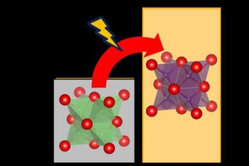
Cost-effective, high-capacity, and cyclable lithium-ion battery cathodes
Charge-recharge cycling of lithium-superrich iron oxide, a cost-effective and high-capacity cathode for new-generation lithium-ion batteries, can be greatly improved by doping with readily available mineral elements. The energy capacity and…
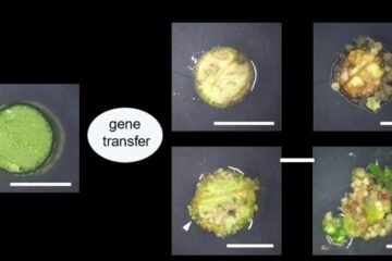
Novel genetic plant regeneration approach
…without the application of phytohormones. Researchers develop a novel plant regeneration approach by modulating the expression of genes that control plant cell differentiation. For ages now, plants have been the…

