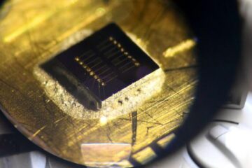
3-D X-ray images of nanoparticles

The device could be used for making better materials, for example for use in electronics, optics and biotechnology.
Transmission electron microscopy (TEM) has traditionally been used to study nanomaterials, but because electrons do not penetrate far into materials, the sample preparation procedure is usually complicated and destructive. Furthermore, TEM only gives two-dimensional images.
The new method shines a powerful X-ray source onto a nanoparticle and collects the X-rays scattered from the sample. Then computers construct a three-dimensional image from that data. The microscope can resolve details down to 17 nanometers, or a few atoms across.
Using the new microscope, Risbud and colleagues were able to take detailed three-dimensional pictures of a “quantum dot” of gallium nitride, and also to study the structure inside it at a nanometer scale. Quantum dots are tiny particles that change their optical and electronic properties, depending on the particle size. Gallium nitride quantum dots could be used in blue-green lasers or flat-panel displays.
“The present work hence opens the door for comprehensive, nondestructive and quantitative 3D imaging of a wide range of samples including porous materials, semiconductors, quantum dots and wires, inorganic nanostructures, granular materials, biomaterials, and cellular structure,” they wrote.
Media Contact
More Information:
http://www.ucdavis.eduAll latest news from the category: Physics and Astronomy
This area deals with the fundamental laws and building blocks of nature and how they interact, the properties and the behavior of matter, and research into space and time and their structures.
innovations-report provides in-depth reports and articles on subjects such as astrophysics, laser technologies, nuclear, quantum, particle and solid-state physics, nanotechnologies, planetary research and findings (Mars, Venus) and developments related to the Hubble Telescope.
Newest articles

Sea slugs inspire highly stretchable biomedical sensor
USC Viterbi School of Engineering researcher Hangbo Zhao presents findings on highly stretchable and customizable microneedles for application in fields including neuroscience, tissue engineering, and wearable bioelectronics. The revolution in…

Twisting and binding matter waves with photons in a cavity
Precisely measuring the energy states of individual atoms has been a historical challenge for physicists due to atomic recoil. When an atom interacts with a photon, the atom “recoils” in…

Nanotubes, nanoparticles, and antibodies detect tiny amounts of fentanyl
New sensor is six orders of magnitude more sensitive than the next best thing. A research team at Pitt led by Alexander Star, a chemistry professor in the Kenneth P. Dietrich…


