Notre Dame imaging specialists create 3-D images to aid surgeons
A paper by the researchers, “3D Printing of Preclinical X-ray Computed Tomographic Data Sets,” was published in the Journal of Visualized Experiments this week.
The strategy was initiated last spring by then-freshman Evan Doney, a Glynn Family Honors student in the laboratory of W. Matthew Leevy, research assistant professor at the Notre Dame Integrated Imaging Facility. “It's a very clever idea,” Leevy says. “He did a lot of it independently. He figured out how to convert the tomographic data to a surface map for editing and subsequent 3D printing.”
The paper reports results based on using X-ray CT data sets from a living Lobund-Wistar rat from the Freimann Life Science Center and from the preserved skull of a New Zealand White Rabbit in the laboratory of Matthew Ravosa. Coauthors of the article with Doney, Leevy, and Ravosa are Lauren Krumdick, Justin Diener, Connor Wathen, Sarah Chapman, Jeremiah Scott and Tony Van Avermaete, all of Notre Dame, and Brian Stamile of MakerBot Industries LLC, a 3-D printing company.
“With proper data collection, surface rendering, and stereolithographic editing, it is now possible and inexpensive to rapidly produce detailed skeletal and soft tissue structures from X-ray CT data,” the paper says. The translation of pre-clinical 3D data to a physical object that is an exact copy of the test subject is a powerful tool for visualization and communication, especially for relating imaging research to students, or those in other fields.”
“Our project with 3-D printing is part of a broader story about 3-D printing in general,” Leevy says, adding that the work has spawned several more ideas and opportunities, such as providing inexpensive models for anatomy students. “There's a market for these bones, both from animals and from humans, and we can create them at incredibly low cost. We're going to explore a lot of these markets.”
A clinical collaborator, Dr. Douglas Liepert from Allied Physicians of Michiana, is enabling the researchers to print non-identifiable human data, expanding the possibilities. “Not only can we print bone structure, but we're starting to collect patient data and print out the anatomical structure of patients with different disease states to aid doctors in surgical preparation,” Leevy says.
Media Contact
More Information:
http://www.nd.eduAll latest news from the category: Medical Engineering
The development of medical equipment, products and technical procedures is characterized by high research and development costs in a variety of fields related to the study of human medicine.
innovations-report provides informative and stimulating reports and articles on topics ranging from imaging processes, cell and tissue techniques, optical techniques, implants, orthopedic aids, clinical and medical office equipment, dialysis systems and x-ray/radiation monitoring devices to endoscopy, ultrasound, surgical techniques, and dental materials.


Newest articles
Economies take off with new airports
A global study by an SUTD researcher in collaboration with scientists from Japan explores the economic benefits of airport investment in emerging economies using nighttime satellite imagery. Be it for…
CAR T–cell immunotherapy targets
Pan-cancer analysis uncovers a new class of promising CAR T–cell immunotherapy targets. Scientists at St. Jude Children’s Research Hospital found 156 potential CAR targets across the brain and solid tumors,…
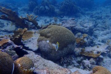
Stony coral tissue loss disease
… is shifting the ecological balance of Caribbean reefs. The outbreak of a deadly disease called stony coral tissue loss disease is destroying susceptible species of coral in the Caribbean…

