


Feeding 9 Billion: Innovations in Agricultural Modeling
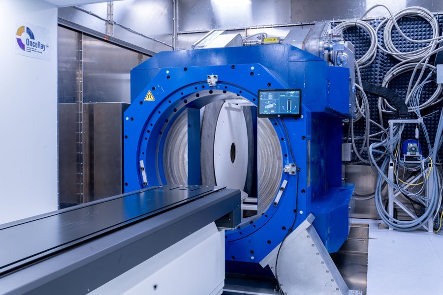
On January 9th, 2024, a scientific prototype for MRI-guided proton therapy was inaugurated in Dresden. With this installation, experts from the fields of medicine, medical physics, biology and engineering are embarking on the scientific testing of a new form of radiotherapy for treating cancer. For the first time globally, a full-body MRI device for real-time imaging is combined with a proton therapy system in the form of a prototype. The inauguration ceremony was held at OncoRay – National Center for Radiation Research in Oncology with Saxony’s Minister-President Michael Kretschmer present. After demonstrating the technical feasibility using a compact MRI device without real-time imaging with a predecessor prototype financed by the Sächsische Aufbaubank in 2019, the Helmholtz-Zentrum Dresden-Rossendorf (HZDR) has now financed the development of a pioneering full-body MRI device with real-time imaging. The infrastructure as well as a number of the personnel are provided by the Dresden University Medical Center.
The aim of the Saxon physicians together with scientists from the HZDR and the Dresden University Medical Center is to monitor cancer patients during their radiation treatment using real-time MRI imaging and thus to significantly improve the targeting accuracy of proton therapy. A globally unique combination of a full-body MRI machine that rotates around the patient for real-time imaging and a proton therapy system was created in Dresden. Scientific operation has now begun in January 2024.
The MRI’s advantage over conventional imaging modalities is that it can visualize the tumor in higher contrast. This makes it possible to better delineate the tumor from surrounding healthy tissue and to define the volume to be irradiated more accurately. Furthermore, MRI imaging can visualize any potential changes in the shape and size of the volume to be irradiated between consecutive radiation sessions. This enables the beam application to be adjusted individually and immediately. In addition, it allows the real-time MRI imaging to visualize tumor movement during a radiation session and to synchronize it with the radiation application. The prototype that has now been installed will be the first of its kind globally to investigate the extent to which the accuracy of proton therapy can be improved with the help of full-body real-time MRI imaging.
At the OncoRay – National Center for Radiation Research in Oncology, the “Experimental MR-Integrated Proton Therapy” research group led by Prof. Aswin Hoffman has developed the new system. This was a technological challenge, as both the MRI device and the proton radiation system work with magnetic fields, which interact with one another and thus influence the quality of the imaging as well as the proton beam application. Having already demonstrated the technical feasibility of simultaneous radiation and imaging using a prior prototype, the research group can now utilize the new system for the first time worldwide to examine the extent to which it is possible to do so using real-time MRI imaging. “This new prototype with integrated full-body MRI makes it possible to visualize moving tumors using high-contrast real-time imaging. Our work aims to develop a technique to irradiate tumors only when they are hit reliably by the proton beam,” says Hoffmann. “The MRI device, which can rotate around the patient, enables us to use innovative types of patient positioning for proton therapy in both lying or in upright positions.” The prototype will be used in future studies to demonstrate the added value of this new prototype for mobile tumors in the chest, abdomen and pelvis.
The prototype has been erected in the proton therapy facility’s experimental room at OncoRay on the premises of the School of Medicine Dresden. There, an international team of researchers is working on innovative ways of treating cancer. Thanks to a large experimental room adjacent to the actual treatment room for patients, the Dresden proton therapy facility enables this unique research work to advance in an interdisciplinary team.
The prototype’s development and installation were made possible in close cooperation with international industry partners. ASG Superconductors in Genoa, Italy manufactured the MRI device, while MagnetTx Oncology Solutions in Edmonton, Canada designed the rotating gantry. Company representatives from both manufacturers were present at the launch ceremony.
Michael Kretschmer, Minister-President of Saxony: “The inauguration of this globally unique combination of an MRI machine with a proton therapy facility marks a milestone for Saxony as a center for science. It is an example of the potential and knowledge available here. At the same time, it becomes clear how we can achieve great things together through strong international collaborations. It is positive and right that the Free State of Saxony specifically supports innovative projects such as this one at the university hospital.”
Sebastian Gemkow, Saxon State Minister for Science: “In order to improve therapies in the long term and to exploit the potential of modern technology, innovative science and research are needed. If, as is the case here, different technological disciplines are able to work closely together with medical professionals, this is an enormous advantage and at the same time a great benefit. The new prototype proves that even the impossible is possible. That is something we can be proud of together.”
Prof. Michael Albrecht, University Hospital Dresden, Medical Director: “What is special about the Dresden University Medical Center is the close connection between patient care and science. We have always placed a particular focus on innovative, modern, therapeutic approaches — especially in oncology. The innovative leap that we are now making with the new prototype demonstrates that we are always at the forefront. A role we can only assume thanks to the positive collaboration with our scientific and industrial partners. This is the only way flagship projects like this are possible.”
Prof. Sebastian M. Schmidt, Helmholtz-Zentrum Dresden-Rossendorf, Scientific Director: “Radiation-based cancer research is one of the HZDR’s major fields of research. We conduct research for healing. Our particular concern is to quickly set research findings into practice for the benefit of patients. We are fulfilling this aspiration precisely with the new prototype.”
Prof. Esther Troost, Dean of the School of Medicine Dresden and Director of the Clinic and Polyclinic for Radiotherapy and Radiation Oncology: “The research alliance of OncoRay – National Center for Radiation Research in Oncology – sees itself as a cross-institutional research platform for medical radiation research in Dresden with a particular focus on translational research. The prototype that has now been inaugurated fits perfectly into this special research environment. Our research goal is to improve cancer treatment through biologically individualized and technologically optimized radiotherapy. We also hope to achieve such improvement with the world’s first MRI-guided proton therapy developed here.”
The prototype’s development and installation were made possible in close cooperation with international industry partners. ASG Superconductors in Genoa, Italy manufactured the MRI device, while MagnetTx Oncology Solutions in Edmonton, Canada designed the rotating gantry. Company representatives from both manufacturers were present at the launch ceremony.
Prof. Aswin Hoffmann
Institute for Radiooncology – OncoRay at the HZDR
Tel.: +49 351 458 3932
aswin.hoffmann@hzdr.de
Simon Schmitt
HZDR Press Officer
Tel.: +49 351 260 3400
s.schmitt@hzdr.de
Annechristin Bonß
University Hospital Dresden, Press Office
Tel.: +49 351 458 4162
pressestelle@ukdd.de


Researchers at Linköping University have successfully visualized blood flow in an artificial heart in real time using magnetic resonance imaging (MRI). The findings, published in Scientific Reports, pave the way for designing artificial hearts that lower the risk of blood clots and red blood cell damage—two of the most common complications in current devices. The project was carried out in collaboration with Scandinavian Real Heart AB, a company working on the development of an artificial heart. “The heart is a…
Around one-third of the 50 million people living with epilepsy worldwide do not respond to anti-seizure medications, leaving them with limited treatment options. Surgical removal of the seizure-causing region can sometimes help, but it is not viable when seizures originate from multiple or unclear brain regions. Deep brain stimulation (DBS) has emerged as a promising alternative for these patients. DBS involves implanting electrodes that deliver controlled electrical impulses to specific brain regions to help control seizures. While stimulation of the…
Each year in the UK, more than 40,000 people suffer an out-of-hospital cardiac arrest (OHCA), yet fewer than 10% survive. Rapid CPR and early use of an Automated External Defibrillator (AED) can at least double survival chances, but in practice, AEDs are often difficult to locate quickly. To address this, researchers at the University of Warwick have partnered with the Welsh Ambulance Services University NHS Trust and drone specialists SkyBound to test whether drones could deliver AEDs directly to the…
The precise excision of tumours is a key and formidable part of oncological surgery. For example, up to 35% of surgical operations for breast cancer result in positive margins, which show that there are cancerous cells in the area around the removed tissue. This increases the risk of cancer recurrence and often leads to repeated surgeries. Before surgery, imaging techniques like ultrasonography are helpful, but during the process, they frequently fail to clearly define the boundaries of the tumour. The…
