Accurate evaluation of chondral injuries by near infrared spectroscopy

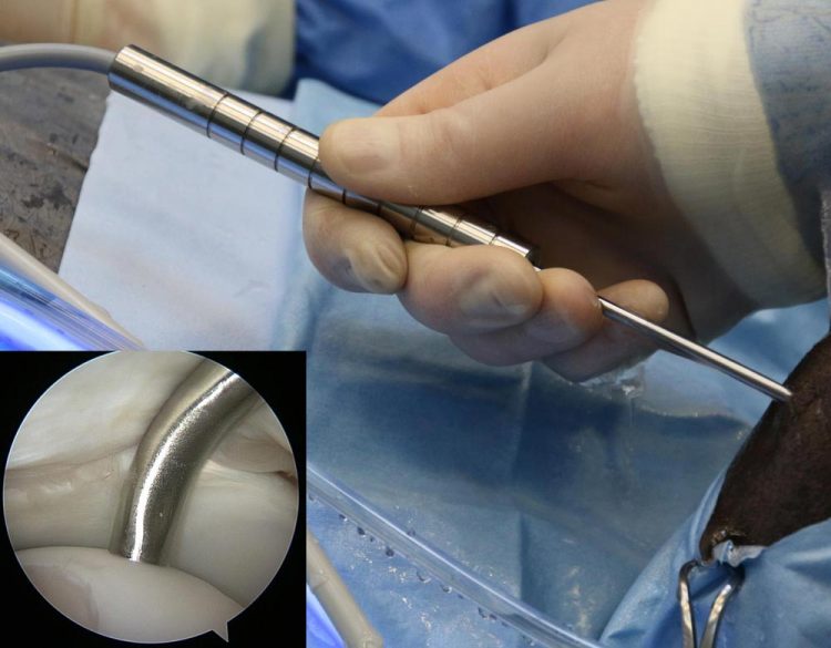
The novel arthroscopic probe in an equine knee joint in vivo with the probe tip in contact with cartilage surface (inset). Credit: Jaakko Sarin
Although no cure currently exists for osteoarthritis, early detection of cartilage lesions could enable halting the disease progression by pharmacological or surgical means. Conventionally, joint health is diagnosed based on patients' symptoms, joint mobility and, if required, with x-ray and magnetic resonance imaging.
Based on these examinations, joint repair surgery may be performed during arthroscopy. The decision on the optimal treatment option is made during the surgery, in which the joint health is evaluated visually and by palpating the cartilage surface with a metallic hook. These techniques are subjective and dependent on surgeons' experience and can, therefore, influence the treatment outcome.
An arthroscopic near infrared spectroscopic probe for evaluation of articular cartilage and subchondral bone structure and composition was developed as part of a PhD thesis at the University of Eastern Finland.
The probe enables enhanced detection of cartilage injuries, as well as evaluation of the integrity of the surrounding tissue. The availability of comprehensive information on the health of joint tissues could substantially enhance the treatment outcome of arthroscopic intervention.
Previously, the near infrared spectroscopy technique has been utilised in, for example, evaluation of grain quality, but its clinical applications are still rare. However, clinical application of the technique is now possible thanks to better availability of computational power along with state-of-the-art mathematical modelling methods, such as neural networks.
With these methods, the relationship between the absorption of near infrared light and tissue properties can be determined. This enables reliable determination of articular cartilage stiffness and subchondral bone mineral density — changes in these tissue properties are prognostic indicators of osteoarthritis.
Since near infrared spectroscopy is not optimal for imaging of tissues, arthroscopically applicable imaging techniques, such as optical coherence tomography and ultrasound imaging, were also used in the study. These techniques have been previously applied in intravascular imaging via specialized 1 mm diameter catheters, which are therefore well-suited for imaging narrow joint cavities. The study compared the reliability of these techniques for evaluation of chondral injuries with that of conventional arthroscopic evaluation.
“Optical coherence tomography was superior to conventional arthroscopy and ultrasound imaging. In contrast to conventional arthroscopic evaluation, optical coherence tomography and ultrasound imaging provide information on inner structures of cartilage and enable, for example, detection of cartilage detachment from subchondral bone,” Researcher Jaakko Sarin from the University of Eastern Finland explains.
###
The doctoral dissertation, entitled Evaluation of chondral injuries using near infrared spectroscopy, is available for download at http://epublications.
The findings were originally reported in Osteoarthritis and Cartilage, Annals of Biomedical Engineering, and Scientific Reports.
For further information, please contact:
Researcher Jaakko Sarin, tel. +358 40 767 7216, jaakko.sarin@uef.fi
Open access article:
Arthroscopic near infrared spectroscopy enables simultaneous quantitative evaluation of articular cartilage and subchondral bone in vivo. Jaakko K. Sarin, Nikae C. R. te Moller,Irina A. D. Mancini, Harold Brommer, Jetze Visser, Jos Malda, P. René van Weeren, Isaac O. Afara & Juha Töyräs. Scientific Reports, volume 8, Article number: 13409 (2018). https:/
Media Contact
All latest news from the category: Medical Engineering
The development of medical equipment, products and technical procedures is characterized by high research and development costs in a variety of fields related to the study of human medicine.
innovations-report provides informative and stimulating reports and articles on topics ranging from imaging processes, cell and tissue techniques, optical techniques, implants, orthopedic aids, clinical and medical office equipment, dialysis systems and x-ray/radiation monitoring devices to endoscopy, ultrasound, surgical techniques, and dental materials.
Newest articles

Superradiant atoms could push the boundaries of how precisely time can be measured
Superradiant atoms can help us measure time more precisely than ever. In a new study, researchers from the University of Copenhagen present a new method for measuring the time interval,…

Ion thermoelectric conversion devices for near room temperature
The electrode sheet of the thermoelectric device consists of ionic hydrogel, which is sandwiched between the electrodes to form, and the Prussian blue on the electrode undergoes a redox reaction…

Zap Energy achieves 37-million-degree temperatures in a compact device
New publication reports record electron temperatures for a small-scale, sheared-flow-stabilized Z-pinch fusion device. In the nine decades since humans first produced fusion reactions, only a few fusion technologies have demonstrated…


