


Feeding 9 Billion: Innovations in Agricultural Modeling
AI Generated Image
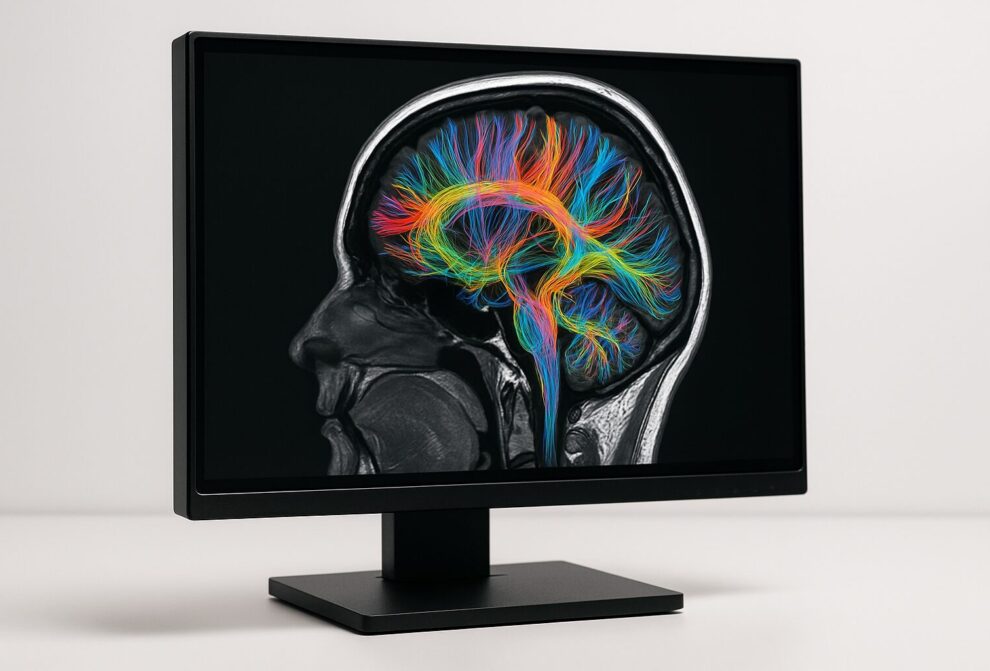
A Japanese research team has demonstrated a new, more accurate way to analyze brain imaging data in children with attention deficit/hyperactivity disorder (ADHD), offering fresh insights into the brain structure differences that underlie the condition. The findings, published in Molecular Psychiatry, could pave the way for earlier diagnosis and more effective, personalized treatments for affected children.
ADHD affects more than 5% of children worldwide, leading to difficulties with attention, hyperactivity, and impulsivity. Brain imaging studies using magnetic resonance imaging (MRI) have long sought to uncover the neurological basis of the disorder, but past results have been mixed—some suggesting reduced gray matter volume (GMV) in children with ADHD, others reporting no difference or even increases.
These inconsistencies often stem from small sample sizes and differences in MRI machines across sites. Previous correction methods, such as ComBat harmonization, help reduce site-related bias but risk “overcorrecting,” removing not only measurement errors but also meaningful biological information.
To overcome these challenges, the researchers applied the traveling-subject (TS) method, which directly accounts for machine-related variation by scanning the same individuals on multiple MRI machines.
In this study, 14 healthy participants were scanned on four different machines over three months to identify measurement biases. This correction model was then applied to an independent dataset from the Child Developmental MRI (CDM) database, which includes MRI data from over 1,000 children. The analyzed sample included 178 typically developing (TD) children and 116 children with ADHD.
“Patients with ADHD displayed smaller brain volumes in regions that are essential for cognitive function and emotional control,” explained Associate Professor Yoshifumi Mizuno of the University of Fukui. “These findings help us better understand why children with ADHD experience such difficulties in daily life.”
By reliably identifying structural brain patterns linked to ADHD, the TS method could improve early detection and monitoring of treatment outcomes.
“Applying the TS harmonization method allows us to more accurately identify brain structure characteristics in ADHD,” said Assistant Professor Qiulu Shou, who led the study. “In the long term, this approach may improve quality of life for affected children and reduce the risk of secondary psychiatric disorders.”
Original Publication
Authors: Qiulu Shou, Masatoshi Yamashita, Yoshiyuki Hirano, Akiko Yao, Min Li, Yide Wang, Yoko Kato, Tokiko Yoshida, Koji Matsumoto, Tetsuya Tsujikawa, Hidehiko Okazawa, Akemi Tomoda, Kuriko Kagitani-Shimono and Yoshifumi Mizuno.
Journal: Molecular Psychiatry
DOI: 10.1038/s41380-025-03142-6
Method of Research: Imaging analysis
Subject of Research: People
Article Title: Brain structure characteristics in children with attention deficit/hyperactivity disorder elucidated using traveling-subject harmonization
Article Publication Date: 8-Aug-2025
COI Statement: The authors declare no biomedical financial interests or potential conflicts of interest.
Original Source: https://www.u-fukui.ac.jp/en-research/108749/
The study aimed to address measurement bias in MRI scans by comparing data from different machines and ensuring accurate brain structure analysis in participants with ADHD and typically developing children.
The researchers used two methods, the TS harmonization method and ComBat harmonization, to estimate and correct measurement bias from different MRI machines, ensuring that the brain data was comparable across all participants.
The study found that after correcting for biases, there were significant differences in brain structures between children with ADHD and typically developing children, indicating that ADHD may be associated with specific structural brain variations.


Scientists uncover how support cells, once thought harmless, send damaging signals that weaken the heart Heart failure (HF) is one of the leading causes of death and disability worldwide, affecting millions of people and placing an enormous burden on healthcare systems. The disease occurs when the heart can no longer pump blood efficiently, leaving patients short of breath, fatigued, and at risk of life-threatening complications. For decades, scientists have focused on studying cardiomyocytes—the heart’s muscle cells responsible for pumping blood—believing…
European research team presents comprehensive review in Brain Medicine on tDCS, rTMS, and DBS for obsessive-compulsive disorder Lausanne, Switzerland – 28 October 2025. In a peer-reviewed article published today in Brain Medicine, a European research team presents a focused review of emerging neuromodulation techniques for treatment-resistant obsessive-compulsive disorder (OCD). The article, “Neuromodulation techniques in obsessive-compulsive disorder: Current state of the art,” examines how transcranial direct current stimulation (tDCS), repetitive transcranial magnetic stimulation (rTMS), and deep brain stimulation (DBS) are changing…
Lawsonia inermis, widely recognised as the source of henna dye used for colouring skin and fabrics, may soon have a life-saving medical application. Researchers at Osaka Metropolitan University have discovered that pigments derived from the plant could help combat liver fibrosis — a serious disease that leads to excessive scar tissue formation in the liver due to chronic injury. Understanding Liver Fibrosis Liver fibrosis occurs when prolonged liver damage — often from factors like alcohol abuse or unhealthy lifestyles —…
Study finds the molecule CCL5 is both protective and harmful, suggesting future drugs could target only its damaging effects Chronic kidney disease (CKD) is a progressive condition in which the kidneys gradually lose their ability to filter waste from the blood. It is a common health concern that affects an estimated 8–16% of the global population, particularly among older adults. CKD can arise from various causes, including glomerulonephritis, a group of diseases that damage the glomeruli, the tiny filtering units…
