


Feeding 9 Billion: Innovations in Agricultural Modeling
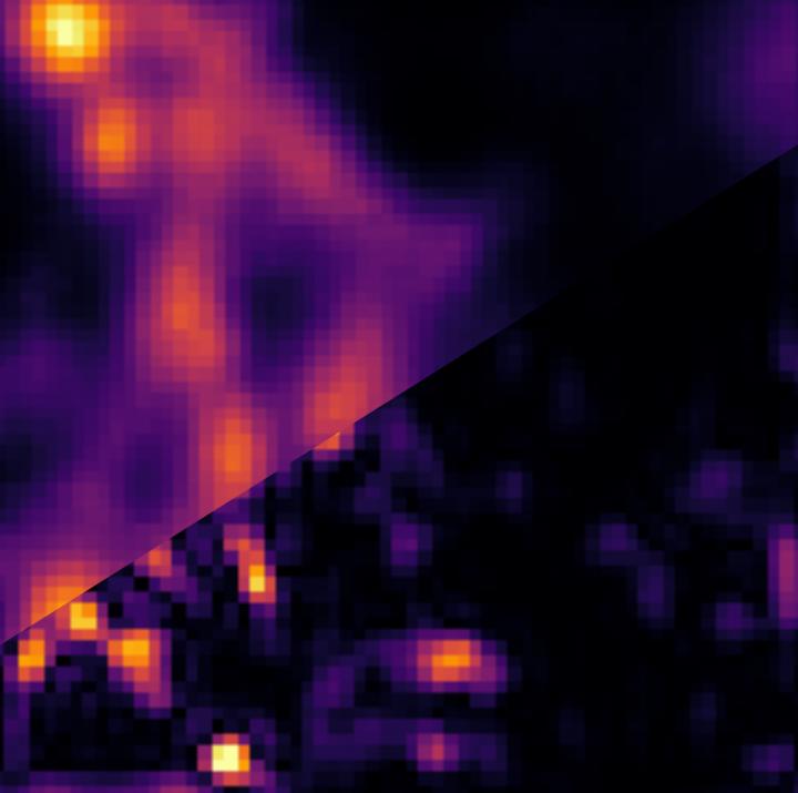
Image of microtubules in a fixed cell sample. A 3 microns x 3 microns confocal scan of microtubules in a fixed 3T3 cell labelled with quantum dots analyzed in two ways. Upper left: image scanning microscopy (ISM), lower right: super-resolution optical fluctuation image scanning microscopy (SOFISM) after Fourier-reweighting. (Source: UW Physics, A. Makowski).
The Polish-Israeli team from the Faculty of Physics of the University of Warsaw and the Weizmann Institute of Science has made another significant achievement in fluorescent microscopy. In the pages of the Optica journal the team presented a new method of microscopy which, in theory, has no resolution limit. In practice, the team managed to demonstrate a fourfold improvement over the diffraction limit.
The continued development of biological sciences and medicine requires the ability to examine smaller and smaller objects. Scientists need to see into the structure of, and the mutual relationships between, for example, proteins in cells. At the same time, the samples being observed should not differ from the structures naturally occurring in biological organisms, which rules out the use of aggressive procedures and reagents. Although it revolutionised the natural sciences, the classical optical microscope is clearly insufficient today. Due to the wavelike nature of light, an optical microscope does not allow imaging structures smaller than about 250 nanometres. As a result, objects closer to each other than half the wavelength of light (which is about 250 nm for green light) cannot be discerned. This phenomenon, known as the diffraction limit, one of the main obstacles in observing the tiniest biological structures, scientists have long attempted to overcome. Electron microscopes provide orders of magnitude better resolution but only allow the examination of inanimate objects, which must be placed in a vacuum and bombarded by an electron beam. For this reason, electron microscopy cannot be used for studying living organisms and the natural processes occurring in them. This is where fluorescence microscopy steps in, hence the rapid development of super-resolution fluorescence microscopy as a field of physical sciences and the two Nobel Prizes already awarded for related research – in 2008 and 2014.
Nowadays several techniques of fluorescence microscopy are available, and some of them have become widespread in biological imaging. Some methods, such as PALM, STORM or STED microscopy, are characterised by an ultra-high resolution and allow discerning objects located just a dozen or so nanometres from each other. However, these techniques require long exposure times and a complex procedure of biological specimen preparation. Other techniques, such as SIM or ISM microscopy, are easy to use, but offer a very limited resolution improvement, allowing to identify structures only half the size of the diffraction limit.
Aleksandra Sroda, Adrian Makowski and Dr. Radek Lapkiewicz from the Quantum Optics Lab at the Faculty of Physics of the University of Warsaw, in cooperation with Prof. Dan Oron’s team from the Weizmann Institute of Science in Israel, have introduced a new technique of super-resolution microscopy, called Super-resolution optical fluctuation image scanning microscopy (SOFISM). In SOFISM, the naturally occurring fluctuations in emission intensity of fluorescent markers are used to further enhance the spatial resolution of an image scanning microscope (ISM). ISM, an emerging super-resolution method, has already been implemented in commercial products and proven valuable for the bio-imaging community. Largely, since it achieves a modest improvement in lateral resolution (x2), with very few changes to the optical setup and without the common handicap of long exposure times. Thus, it enables a natural extension of the capabilities of a standard confocal microscope. ISM uses a confocal microscope in which a single detector is replaced with a detector array. In SOFISM correlations of intensities detected by multiple detectors are computed. In principle, the measurement of the n-th order correlation can lead to a factor of 2n resolution improvement with respect to the diffraction limit. In practice, the resolution achievable for higher-order correlations is limited by the signal-to-noise ratio of the measurements.
“SOFISM is a compromise between ease of use and resolution. We believe that our method will fill the niche between the complex, difficult-to-use techniques providing very high resolution and the easy-to-use lower-resolution methods. SOFISM does not have a theoretical resolution limit, and in our article, we demonstrate results which are four times better than the diffraction limit. We also show that the SOFISM method has a high potential in the imaging of three-dimensional biological structures,” said Dr. Radek Lapkiewicz.
Crucially, SOFISM is, in its technical aspects, highly accessible, as it only requires introducing a small modification to the widely-used confocal microscope – replacing its photomultiplier tube with a SPAD array detector. In addition, it is necessary to slightly increase the measurement time and change the data processing procedure. “Until recently, SPAD array detectors were expensive and their specifications were not sufficient for correlation-based microscopy. This situation has recently changed. The new SPAD detectors introduced last year removed both the technological and price-related barriers. This makes us think that fluorescence microscopy techniques such as SOFISM might, in a few years’ time, become widely used in the field of microscopic examination,” stressed Dr. Lapkiewicz.
The project was carried out under the FIRST TEAM program of the Foundation for Polish Science.
Physics and Astronomy first appeared at the University of Warsaw in 1816, under the then Faculty of Philosophy. In 1825 the Astronomical Observatory was established. Currently, the Faculty of Physics’ Institutes include Experimental Physics, Theoretical Physics, Geophysics, Department of Mathematical Methods and an Astronomical Observatory. Research covers almost all areas of modern physics, on scales from the quantum to the cosmological. The Faculty’s research and teaching staff includes ca. 200 university teachers, of which 87 are employees with the title of professor. The Faculty of Physics, University of Warsaw, is attended by ca. 1000 students and more than 170 doctoral students.
SCIENTIFIC PAPERS:
Aleksandra Sroda, Adrian Makowski, Ron Tenne, Uri Rossman, Gur Lubin, Dan Oron, Radek Lapkiewicz: ”SOFISM: Super-resolution optical fluctuation image scanning microscopy”, Optica Vol. 7, Issue 10, pp. 1308-1316 (2020).
DOI: https:/
CONTACTS:
Dr. Radek Lapkiewicz
Faculty of Physics, University of Warsaw
tel.: +48 508 615 206
email: radek.lapkiewicz@fuw.edu.pl
RELATED LINKS:
http://quantumoptics.
The website of Dr. R. Lapkiewicz’s research group
https:/
The website of prof. Dan Oron’s research group
http://www.
The Faculty of Physics, University of Warsaw website.
https:/
Press office of the Faculty of Physics, University of Warsaw.
IMAGES:
FUW201007b_fot01
https:/
Image of microtubules in a fixed cell sample. A 3 μm x 3 μm confocal scan of microtubules in a fixed 3T3 cell labelled with quantum dots analyzed in two ways. Upper left: image scanning microscopy (ISM), lower right: super-resolution optical fluctuation image scanning microscopy (SOFISM) after Fourier-reweighting. (Source: UW Physics, A. Makowski).


Astronomers have created a galactic masterpiece: an ultra-detailed image that reveals previously unseen features in the Sculptor Galaxy. Using the European Southern Observatory’s Very Large Telescope (ESO’s VLT), they observed this nearby galaxy in thousands of colours simultaneously. By capturing vast amounts of data at every single location, they created a galaxy-wide snapshot of the lives of stars within Sculptor. “Galaxies are incredibly complex systems that we are still struggling to understand,” says ESO researcher Enrico Congiu, who led a…

NASA spacecraft make progress in final commissioning, preliminary science operations SAN ANTONIO — June 10, 2025 — Southwest Research Institute’s Dr. Craig DeForest discussed the latest accomplishments of NASA’s PUNCH (Polarimeter to Unify the Corona and Heliosphere) mission during a media event at the 246th American Astronomical Society meeting in Anchorage, Alaska. As the spacecraft constellation completes commissioning, early PUNCH data showed coronal mass ejections, or CMEs, as they erupted from the Sun and traveled across the inner solar system….

Using advanced computational modelling, a research team led by the University of Oxford, working in partnership with the Instituto Superior Técnico in the University of Lisbon, has achieved the first-ever real-time, three-dimensional simulations of how intense laser beams alter the ‘quantum vacuum’—a state once assumed to be empty, but which quantum physics predicts is full of virtual electron-positron pairs. Excitingly, these simulations recreate a bizarre phenomenon predicted by quantum physics, known as vacuum four-wave mixing. This states that the combined…

An international team of astronomers, including researchers from the University of Liège and collaborators in UK, Chile, the USA, and Europe, has discovered a giant planet orbiting the smallest known star to host such a companion The host star, TOI-6894, is a red dwarf with only 20% the mass of the Sun, typical of the most common stars in our galaxy. Until now, such low-mass stars were not thought capable of forming or retaining giant planets. But as published today…


