


Feeding 9 Billion: Innovations in Agricultural Modeling

3D MRI can detect pancreatic cancer when it is smaller and patients have a greater likelihood of survival, a new study shows.
The study included 57 patients who had clinical symptoms of pancreatic cancer. All had contrast enhanced 3D gradient-echo MRI examinations. Radiologists correctly identified pancreatic cancer in 24 patients, said Richard Semelka, MD, professor of radiology, at the University of North Carolina, Chapel Hill, and an author of the study. Eight of the cancers found were less than two centimeters in size, Dr. Semelka said. “Currently patients with pancreatic cancer are treated with complete surgical resection and the smaller the tumor, the easier it is to remove,” he said.
Pancreatic cancer is usually diagnosed too late, said Dr. Semelka. About 40,000 people are diagnosed with the disease in the U.S. each year, and nearly all of them die. Pancreatic cancer is the fourth most common cause of cancer death in the U.S. “The symptoms of the disease are somewhat nonspecific and can easily be misinterpreted. In addition, the disease is very aggressive so if the disease is missed or the diagnosis is delayed, the patient’s chance for survival is dismal,” he said.
3D MRI did indicate pancreatic cancer in three patients, but biopsy showed they did not have the disease. However, one did have a neuroendocrine tumor and one had focal pancreatitis. Three patients were lost to follow-up, said Dr. Semelka. “No patient with a study interpreted as normal was subsequently found to have pancreatic cancer,” he added.
“We are now working with our internists to detect this disease earlier,” said Dr. Semelka. “We are encouraging them to refer their patients for a 3D MRI examination if a patient has severe mid-abdominal pain not explained by a back problem, sudden development of diabetes and/or sudden development of jaundice. Radiologists who are reading 3D MR images of patients with abdominal pain should also look for pancreatic cancer even if the patient didn’t have the examination for that purpose,” he said.
The study appears in the September 2005 issue of the American Journal of Roentgenology.


A Kobe University team was able to edit the DNA of Lactobacillus strains directly without a template from other organisms. This technique is indistinguishable from natural variation and enabled the researchers to create a strain that doesn’t produce diabetes-aggravating chemicals. Humans have improved the microorganisms we rely on for millennia, selecting variants that are better able to produce wine, yogurt, natto and many other products. More recently, direct genetic modification has emerged as a tool to exert more precise and…
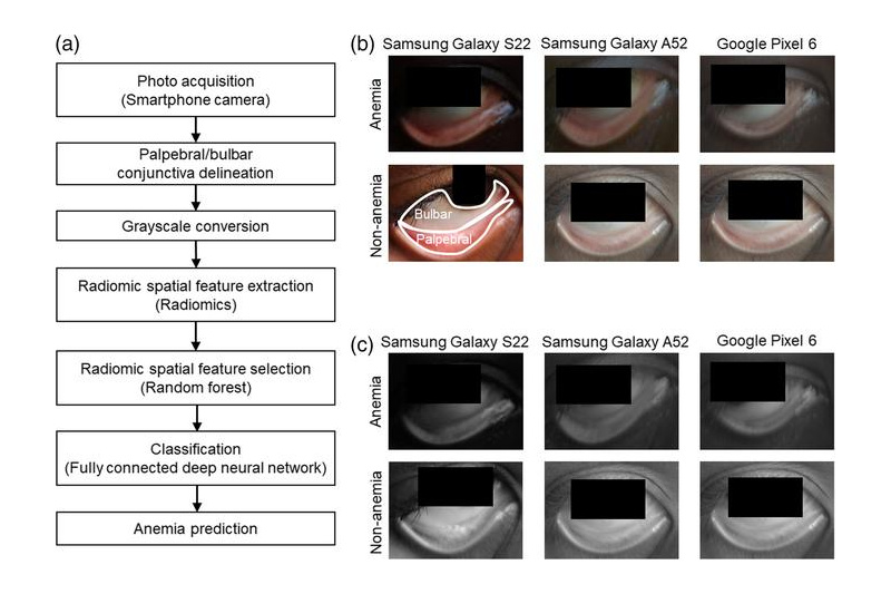
Noninvasive method detects anemia in children by analyzing smartphone photos of the eye’s conjunctiva Anemia, a condition marked by low levels of hemoglobin in the blood, affects nearly 2 billion people worldwide. Among them, school-age children in low- and middle-income countries are particularly vulnerable. Left untreated, anemia in children can interfere with growth, learning, and overall development. Detecting the condition early is essential, but standard diagnostic methods require blood samples and lab equipment—resources that are often unavailable in low-income areas….

Designed by University of Missouri researchers, the device includes AI technology to detect potential heart problems with over 90% accuracy, making it a promising tool for at-home monitoring When we move, it’s harder for existing wearable devices to accurately track our heart activity. But University of Missouri researchers found that a starfish’s five-arm shape helps solve this problem. Inspired by how a starfish flips itself over — shrinking one of its arms and using the others in a coordinated motion…

A pan-Canadian team has developed a new way to quickly find personalized treatments for young cancer patients, by growing their tumours in chicken eggs and analyzing their proteins. The team, led by researchers from the University of British Columbia and BC Children’s Hospital Research Institute, is the first in Canada to combine these two techniques to identify and test a drug for a young patient’s tumour in time for their treatment. Their success in finding a new drug for the…


