


Feeding 9 Billion: Innovations in Agricultural Modeling
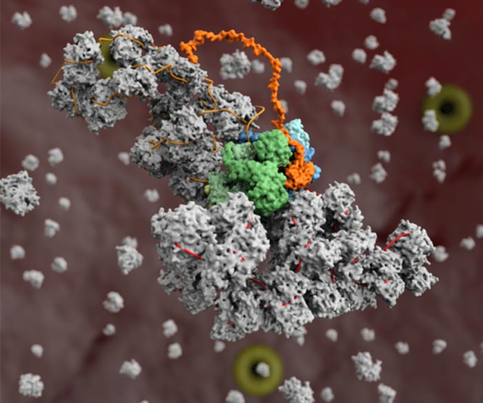
A model of the influenza virus replication machinery in the process of the viral RNA polymerase (blue) copying a viral RNA genome segment (red) in complex with a second copy of the polymerase (green) and host protein ANP32A (orange), and the assembly of the newly synthesised RNA segment (orange) into a ribonucleoprotein complex (grey).
Credit: Diamond Light Source / Trials in Microbiology
Structural insights reveal new potential drug targets for the development of novel antiviral drugs to inhibit influenza virus replication.
A team of scientists at University of Oxford have worked with multiple techniques at Diamond Light Source, the UK’s national synchrotron, to solve the structure of the influenza replication machinery and to determine how it interacts with cellular proteins. This new research furthers understanding of influenza replication and how the virus adapts to different hosts. These structural insights have revealed new potential drug targets for the development of novel antiviral drugs to inhibit influenza virus replication.
The paper “A structural understanding of influenza virus genome replication.” Trends in Microbiology (2022). DOI: 10.1016/j.tim.2022.09.015 outlines the findings generated using X-ray crystallography and small angle X-ray scattering (SAXS) at the synchrotron and cryo-Electron Microscopy (cryo-EM) at Diamond’s electron Bio-Imaging Centre (eBIC). The paper will appear in print in the March 2023 issue with the image on its front cover of the influenza virus replication machinery.
Beyond causing seasonal flu, influenza can become pandemic when it jumps from animals to humans. By taking a closer look at the replication cycle of the virus, the researchers are piecing together how influenza hijacks human and animal cells for its replication. This research is crucial to understanding how a cellular protein (ANP32A) partly accounts for the host-jumping barrier. By studying which regions of the viral polymerase interact with ANP32A, the researchers determined that a mutation in avian influenza’s polymerase may allow it to interact with human ANP32A, permitting that strain of bird flu to jump into human hosts2.
The influenza virus stores its genes in RNA and the virus synthesises its own RNA polymerase to replicate its genome. This viral polymerase has multiple functions in addition to replication, which collaborative research across Diamond has helped to elucidate. These studies show that the polymerase regulates the timing of transcription — the first step in protein synthesis — and replication, which can only begin once viral proteins have been produced. The findings reveal how the polymerase interacts with a cellular protein, ANP32A, and appropriates it to shelter viral RNA from detection by the immune system.
The currently circulating influenza A viruses are thought to be the evolutionary progeny of the virus that caused the 1918–1919 global pandemic, which was responsible for between 50 and 100 million deaths worldwide. Influenza viruses are normally restricted to infecting one type of animal host, such as birds, and require specific adaptations to jump to a different animal, like humans. The 1918 influenza virus is thought to have jumped from waterfowl into humans and is considered the ‘founder virus’ that has contributed viral genome segments for all subsequent epidemic and pandemic strains. In a study published earlier this year, the group determined structures of the polymerase from the 1918 pandemic influenza virus and identified sites on the surface of the polymerase that are sensitive to inhibition1. This in turn can help identify and validate targets for drug discovery.
This research is crucial to understanding how ANP32A partly accounts for the host-jumping barrier. ANP32A differs greatly between humans and birds, forcing animal and avian influenza viruses to evolve to become less alike. Structural biology research at Diamond provides insights into the pandemic potential of different strains of flu. By studying which regions of the viral polymerase interact with ANP32A, the researchers determined that a mutation in avian influenza’s polymerase may allow it to interact with human ANP32A, permitting that strain of bird flu to jump into human hosts2.
Structural characterisation of large protein complexes is a challenge, and the influenza replication complex was no exception. X-ray crystallography at beamlines I03 and I24 was used to determine the structure of the viral polymerase in near-atomic detail, revealing that the single polymerases pair together to form dimers. To complement the crystal structure of the dimers, a structural technique known as SAXS was performed in solution at beamline B21 to demonstrate the importance of dimer formation to the function of the polymerases.
The researchers proposed that single RNA polymerases carry out transcription early in infection and switch to replication later only when they couple together as dimers, following the production of additional copies of the polymerase3.
To expand on this structural work further, the research team performed cryo-EM at eBIC. Professor Jonathan Grimes at University of Oxford explains: “Cryo-EM has allowed us to begin to look at very interesting protein complexes that we would find impossible to grow crystals of in the lab.”
Interactions between RNA and the viral polymerase were determined using cryo-EM, revealing that one polymerase in the dimer replicates the viral genome while the other coats the newly formed RNA in viral proteins that shelter it from immune sensors. Intriguingly, influenza hijacks the cellular protein ANP32A to stabilise the dimers and to assist with coating and hiding viral RNA from immune detection.
“Diamond democratises science,” explains Grimes. “The fact that all of these techniques exist in one place and are available to the scientific community is a hugely valuable resource. These world class cutting-edge facilities are freely available to scientists from universities and institutes across the UK and EU with interesting and important biological questions.”
Corresponding author on the Trends in Microbiology review, Professor Ervin Fodor, University of Oxford, concludes: “These studies help us to identify and validate targets for drug discovery. We are hoping that the new insights generated into workings of the influenza virus transcription machinery using the technologies at Diamond will eventually lead to novel antivirals targeting the influenza polymerase.”
References
Related publication
Zhu Z. et al. A structural understanding of influenza virus genome replication. Trends in Microbiology (2022). DOI: 10.1016/j.tim.2022.09.015
IMAGE
A model of the influenza virus replication machinery in the process of the viral RNA polymerase (blue) copying a viral RNA genome segment (red) in complex with a second copy of the polymerase (green) and host protein ANP32A (orange), and the assembly of the newly synthesised RNA segment (orange) into a ribonucleoprotein complex (grey).
[VIDEO – https://vimeo.com/diamondlightsource/fluanimation
This animation explores how the flu virus interacts with our bodies, focusing on a protein (ANP32A) that aids replication of the virus. Knowing the structure and function of this stabilising protein will enable scientists to work on new treatments to fight the disease.
ENDS
For further information please contact Diamond Communications: Lorna Campbell +44 7836 625999 or Isabelle Boscaro-Clarke +44 1235 778130 Diamond Light Source: www.diamond.ac.uk Twitter: @DiamondLightSou
Diamond Light Source provides industrial and academic user communities with access to state-of-the-art analytical tools to enable world-changing science. Shaped like a huge ring, it works like a giant microscope, accelerating electrons to near light speeds, to produce a light 10 billion times brighter than the Sun, which is then directed off into 33 laboratories known as ‘beamlines’. In addition to these, Diamond offers access to several integrated laboratories including the world-class Electron Bio-imaging Centre (eBIC) and the Electron Physical Science Imaging Centre (ePSIC).
Diamond serves as an agent of change, addressing 21st century challenges such as disease, clean energy, food security and more. Since operations started, more than 14,000 researchers from both academia and industry have used Diamond to conduct experiments, with the support of approximately 760 world-class staff. Almost 12,000 scientific articles have been published by our users and scientists.
Funded by the UK Government through the Science and Technology Facilities Council (STFC), and by the Wellcome Trust, Diamond is one of the most advanced scientific facilities in the world, and its pioneering capabilities are helping to keep the UK at the forefront of scientific research.
Diamond was set-up as an independent not for profit company through a joint venture, between the UKRI’s Science and Technology Facilities Council and one of the world’s largest biomedical charities, the Wellcome Trust – each respectively owning 86% and 14% of the shareholding.
Journal: Trends in Microbiology
DOI: 10.1016/j.tim.2022.09.015
Method of Research: Imaging analysis
Subject of Research: Cells
Article Title: A structural understanding of influenza virus genome replication
Article Publication Date: 3-Nov-2022
Media Contact
Lorna Arthur
Diamond Light Source
lorna.campbell@diamond.ac.uk
Office: 44-783-662-5999


A recent study published in the Annals of the Entomological Society of America provides the most comprehensive insight to date into the development of this atypical fly and its live birth—a uncommon occurrence among flies. Undergraduate student Parker Henderson ‘22 from St. Olaf College spearheaded the project, which unveiled significant findings regarding the reproductive biology of Ormia ochracea, a parasitic fly renowned for its hyperacute directional hearing that enables it to locate chirping crickets. The scientists utilised dissection, fluorescence labelling,…

Researchers at Osaka Metropolitan University have elucidated a longstanding enigma in sonochemistry: the reason chemical reactions decelerate when ultrasonic power is very high. Their discoveries facilitate more intelligent use of ultrasound in scientific and industrial contexts, including environmental remediation and the synthesis of beneficial nanoparticles. Science Behind Ultrasound and Chemical Reactions Despite being imperceptible to the human ear, ultrasonography significantly influences sonochemistry. Ultrasonic waves applied to a liquid produce small bubbles that swiftly expand and disintegrate, a phenomenon known as…

Quantum computers encounter a significant obstacle in their pursuit of practical applications: their constrained capacity to rectify emerging computational mistakes. To create genuinely dependable quantum computers, researchers must replicate quantum calculations on classical computers to validate their accuracy – an essential yet exceptionally challenging endeavour. Researchers from Chalmers University of Technology in Sweden, the University of Milan, the University of Granada, and the University of Tokyo have introduced a pioneering method for simulating particular forms of error-corrected quantum computations, marking…

The Researchers at ETH Zurich have used nuclear magnetic resonance (NMR) to investigate the atomic surroundings. They have also done it for the spatial orientation of individual platinum atoms embedded in solid supports. This technique is reliable for improving the design and production of single-atom catalysts in the future. Catalysis is the process of accelerating chemical reactions by introducing a specific substance called a catalyst. They are vital to both industry and everyday life. Approximately 80% of all chemical products…


