Observing the internal 3D structure of the nipple to understand and fight breast cancer
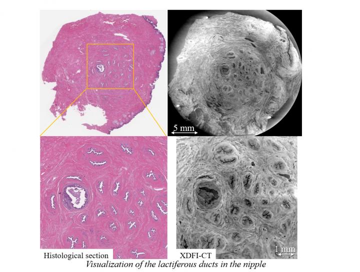
Visualization of the lactiferous ducts in the nipple Credit: Naoki Sunaguchi, Nagoya University
Conventional reconstruction from multiple two-dimensional tissue slices is a laborious process, is invasive, and does not give a clear understanding of the 3D structure of the nipple. Thus, the search for better imaging techniques continues.
Recently, Associate Professor Sunaguchi from Nagoya University and his colleagues saw the potential of X-ray dark-field computed tomography (or XDFI-CT) as a promising alternative to existing nipple-exploration methods, such as needle biopsy or ductoscopy.
XDFI-CT is a technique that can create a 3D rendering of a structure based on the distribution of its electron density and refractive index. Sunaguchi and team hypothesized that XDFI-CT could provide a 3D reconstruction of the human nipple that is at least as precise as images built from 2D slices, with the added benefit that the proposed approach leaves the nipple intact and therefore has wider applications.
“In this study, which has been published in Breast Cancer Research and Treatment, we examined 51 human nipples by XDFI-CT and visualized the 3D arrangements of the nipple ducts,” explains Assoc. Prof. Sunaguchi. First, XDFI-CT was used to obtain 2D sections of the nipple so as to compare them with conventional tissue sections observed through light microscopy.
The results showed that the 2D CT slices were structurally compatible with the ones obtained through the conventional approach, giving a first demonstration of the capabilities of the method.
The CT-derived 2D slices were afterwards easily turned into full 3D renderings of the nipples, allowing the researchers to count the number of milk ducts and analyze the overall ductal structure, which varied from patient to patient. “Using 3D volume renderings of the 51 samples, we discovered three different types of duct arrangements,” comments Assoc. Prof. Sunaguchi.
These three types of arrangements were “convergent,” in which the ducts converged in a bowl shape centered at the tip of the nipple; “straight.” where the ducts grew parallelly from the base to the tip; and “divergent,” where the ducts diverged as they came closer to the tip.
The findings of this study also provided evidence for “sick lobe theory,” which postulates that non-invasive ductal carcinoma (DCIS) begins in a single mammary lobe. In one particular sample, the researchers noted that three seemingly separate ducts with DCIS converged into a single opening near the tip of the nipple, implying that the three DCIS-ridden ducts were derived from the same cancerous mammary lobe.
Overall, the proposed strategy is very promising not only for analyzing the structure of the nipple but also for scanning other soft tissues or organs. Though there are some technical limitations to currently implementing XDFI-CT for diagnostic or exploratory purposes, the approach employed in the present study will certainly become very relevant once these hurdles are overcome.
###
The article, “Three-dimensional microanatomy of human nipple visualized by X-ray dark-field computed tomography,” was published in the journal Breast Cancer Research and Treatment at DOI: 10.1007/s10549-020-05574-w.
Authors:
Naoki Sunaguchi1, Daisuke Shimao2, Tetsuya Yuasa3, Shu Ichihara4, Rieko Nishimura4, Risa Oshima2, Aya Watanabe1, Kikuko Niwa1 and Masami Ando5
Affiliations:
1Graduate School of Medicine, Nagoya University
2Department of Radiological Technology, Hokkaido University of Science
3Graduate School of Engineering and Science, Yamagata University
4Department of Pathology, Nagoya Medical Center
5Comprehensive Research Organization for Science and Society
For more information, contact:
Naoki Sunaguchi
Associate Professor, Graduate School of Medicine, Nagoya University
Email: sunaguchi@met.nagoya-u.ac.jp
About Nagoya University, Japan
Nagoya University has a history of about 150 years, with its roots in a temporary medical school and hospital established in 1871, and was formally instituted as the last Imperial University of Japan in 1939. Although modest in size compared to the largest universities in Japan, Nagoya University has been pursuing excellence since its founding. Six of the 18 Japanese Nobel Prize-winners since 2000 did all or part of their Nobel Prize-winning work at Nagoya University: four in Physics – Toshihide Maskawa and Makoto Kobayashi in 2008, and Isamu Akasaki and Hiroshi Amano in 2014; and two in Chemistry – Ryoji Noyori in 2001 and Osamu Shimomura in 2008. In mathematics, Shigefumi Mori did his Fields Medal-winning work at the University. A number of other important discoveries have also been made at the University, including the Okazaki DNA Fragments by Reiji and Tsuneko Okazaki in the 1960s; and depletion forces by Sho Asakura and Fumio Oosawa in 1954.
Media Contact
All latest news from the category: Health and Medicine
This subject area encompasses research and studies in the field of human medicine.
Among the wide-ranging list of topics covered here are anesthesiology, anatomy, surgery, human genetics, hygiene and environmental medicine, internal medicine, neurology, pharmacology, physiology, urology and dental medicine.


Newest articles

Properties of new materials for microchips
… can now be measured well. Reseachers of Delft University of Technology demonstrated measuring performance properties of ultrathin silicon membranes. Making ever smaller and more powerful chips requires new ultrathin…

Floating solar’s potential
… to support sustainable development by addressing climate, water, and energy goals holistically. A new study published this week in Nature Energy raises the potential for floating solar photovoltaics (FPV)…

Skyrmions move at record speeds
… a step towards the computing of the future. An international research team led by scientists from the CNRS1 has discovered that the magnetic nanobubbles2 known as skyrmions can be…

