


Feeding 9 Billion: Innovations in Agricultural Modeling
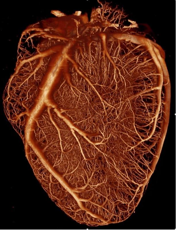
Three-dimensional reconstruction of the blood vessels perfusing the heart muscle
© Nature Reviews Cardiology
Whether patients suffer from acutely or chronically narrowed coronary vessels – quantitative imaging techniques are indispensable when it comes to detecting and treating circulatory disorders of the heart muscle or preventing them in time.
An interdisciplinary team, in which scientists from the German Centre for Cardiovascular Research (DZHK) also played a leading role, has now for the first time determined which method is best suited for patients with different clinical pictures in order to measure the heart’s blood supply.
The researchers have published the results as a joint consensus statement in Nature Reviews Cardiology.
The core of the work is a table with specific recommendations for action, which clearly shows which quantitative imaging methods are appropriate for each patient and the clinical picture at hand.
“What is special about this table is that scientists from different disciplines, namely cardiologists, physiologists, nuclear medicine specialists, physicists and radiologists, have compiled this table for the available imaging methods,” says Professor Marc Dewey, DZHK scientist and Deputy Director of the Department of Radiology at the Mitte campus of Charité – Universitätsmedizin Berlin.
“Until now, there have only been guidelines of individual professional associations or individual techniques have been the focus of attention.”
New findings on the method of choice
The advantages and disadvantages of the different techniques were discussed by the scientists using a systematic, multi-stage evaluation procedure. For some forms of disease, it became clear that one method is superior to all others, while for others, several are possible. “These findings have not been seen before and I hope that they will change clinical practice,” says Dewey.
For example, patients who also suffer from cardiac heart failure benefit most from magnetic resonance imaging (MRI) because, in addition to the blood flow, the heart function can also be assessed – and whether connective tissue has replaced the heart muscle (fibrosis).
Invasive flow measurements are very well suited for patients with known coronary heart disease (CHD) or a high probability of CHD. Positron emission tomography (PET) provides the most accurate absolute quantification and is therefore particularly suitable for patients with CHD of multiple vessels.
Scintigraphy, on the other hand, is the most widely available procedure, and thanks to new technologies, quantification is now also possible. Echocardiography is the method of choice when doctors want to visualize the blood flow in the heart of bedridden patients, as it allows them to perform the examination at the bedside.
Computed tomography (CT) can also measure the blood flow and is the only method that enables the simultaneous analysis of possible coronary stenosis and plaques.
“Our consensus document helps to select the best possible diagnostic strategy and could therefore help to develop individualized suggestions for subsequent therapy,” says Dewey. He also believes that it is important to give patients access to this chart to enable them to bring in their own wishes and ideas.
Open process
The Berlin radiologist expects the recommendations to remain valid for four to five years before they have to be revised in view of the constantly advancing technologies. He and his colleagues are open to new developments and members. So far, their team has been made up of European scientists, but colleagues from Asia or the USA are also welcome.
In this respect, their publication could also be seen as a call for all those who wish to participate in this decision-making process in the future, for instance for getting involved in the next topic, coronary arteries. The second meeting of the Quantitative Cardiac Imaging Study Group on March 9, 2021 in Berlin will be the starting point for this.
Prof. Dr. med. Marc Dewey, Deputy Director of the Department of Radiology at the Mitte Campus of Charité – Universitätsmedizin Berlin, Mitte Campus, marc.dewey(at)charite.de
Original work: Clinical quantitative cardiac imaging for the assessment of myocardial ischaemia
Marc Dewey, Maria Siebes, Marc Kachelrieß, Klaus F. Kofoed, Pál Maurovich-Horvat, Konstantin Nikolaou, Wenjia Bai, Andreas Kofle, Robert Manka, Sebastian Kozerke, Amedeo Chiribiri, Tobias Schaeffter, Florian Michallek, Frank Bengel, Stephan Nekolla, Paul Knaapen, Mark Lubberink, Roxy Senior, Meng-Xing Tang, Jan J. Piek, Tim van de Hoef, Johannes Martens and Laura Schreiber & on behalf of the Quantitative Cardiac Imaging Study Group. Nat Rev Cardiol (2020). DOI:10.1038/s41569-020-0341-8
https://dzhk.de/en/news/latest-news/article/how-well-is-the-heart-perfused-inter…


The idea seems futuristic: At ETH Zurich, various disciplines are working together to combine conventional materials with bacteria, algae and fungi. The common goal: to create living materials that acquire useful properties thanks to the metabolism of microorganisms – “such as the ability to bind CO2 from the air by means of photosynthesis,” says Mark Tibbitt, Professor of Macromolecular Engineering at ETH Zurich. An interdisciplinary research team led by Tibbitt has now turned this vision into reality: it has stably…

About 100 cells divide every second in our body. A key protein in cell division is a protein kinase termed Plk1, because it activates other proteins involved in this process. Plk1 is also overexpressed in many types of cancer. This makes it a promising target for cancer therapies. However, drugs that inhibit Plk1 have often proven ineffective. New findings by researchers led by Peter Lenart and Monica Gobran may help to improve therapeutic approaches. They discovered a previously unknown function…

Good news for the German research fleet, German shipbuilding, and international polar research alike: the new Polarstern will be constructed in Wismar by thyssenkrupp Marine Systems. The company received the official contract to construct a new research icebreaker from the Alfred Wegener Institute (AWI) today, marking the end of a two and a half-year-long Europe-wide call for tenders. The new flagship of German climate research will cost an estimated 1.185 billion euros. Following five years of construction, she is to…

A new radio telescope on the top of Germany’s Zugspitze mountain will help unravel the secrets of the universe. The project is led by the Chair for Astronomy at the University of Würzburg. Thorsten Glauber, State Minister in the Bavarian State Ministry of the Environment and Consumer Protection, spoke of a “new chapter in space research”. And of a further step “to consolidate Bavaria’s place in the premier league of research“. What made the minister so enthusiastic is a state-of-the-art…


