


Feeding 9 Billion: Innovations in Agricultural Modeling
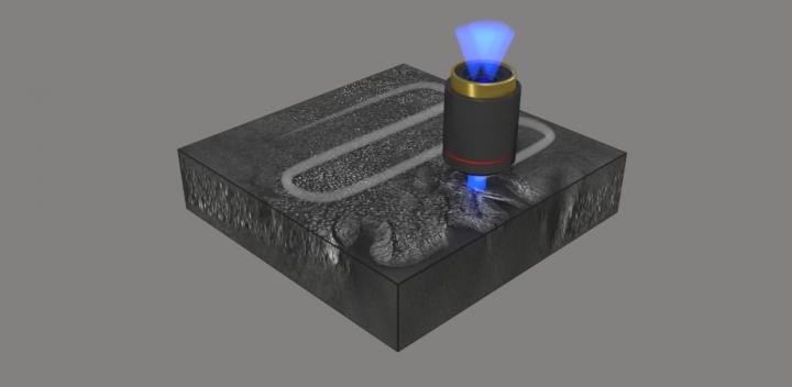
Schematic of the imaging of pathological tissue 3D structure by combining optical diffraction tomography and automated stitching. Image credit: Hugonnet et al., doi 10.1117/1.AP.3.2.026004
Credit: Hugonnet et al.
New optical diffraction tomography method increases imaging speed and resolution for improved diagnostics in histopathology.
Histology is the study of biological tissues at a microscopic level. Also called microscopic anatomy, histology is widely used to provide diagnosis of cancer and other diseases. For example, tissue samples obtained during surgery might help to determine whether further surgical action is needed, and further surgery may be avoided if a diagnosis can be rapidly obtained during an operation.
Traditional methods in histopathology are generally limited to thin specimens and require chemical processing of the tissue to provide sufficiently high contrast for imaging, which slows the process. A recent advance in histopathology eliminates the need for chemical staining and enables high-resolution imaging of thick tissue sections. As reported in Advanced Photonics, an international research team recently demonstrated a 3D label-free quantitative phase imaging technique that uses optical diffraction tomography to obtain volumetric imaging information. Automated stitching simplifies the image acquisition and analysis.
Optical diffraction tomography is a microscopy technique for reconstructing the refractive index of a tissue sample from its scattered field images obtained with various illumination angles. It enables label-free high contrast visualization of transparent samples. The complex scattered field transmitted through the sample is first retrieved using off-axis holography, then the scattered fields obtained with various angle of illuminations are mapped in the Fourier space enabling the reconstruction of the sample refractive index.
A recognized limitation of optical diffraction tomography is due to the complex distribution of refractive indexes, which results in significant optical aberration in the imaging of thick tissue. To overcome this limitation, the team used digital refocusing and automated stitching, enabling volumetric imaging of 100-m-thick tissues over a lateral field of view of 2 mm 1.75 mm while maintaining a high resolution of 170 nm 170 nm 1400 nm. They demonstrated that simultaneous visualization of subcellular and mesoscopic structures in different tissues is enabled by high resolution combined with a wide field of view.
The researchers demonstrated the capacity of their novel method by imaging a variety of different cancer pathologies: pancreatic neuroendocrine tumor, intraepithelial neoplasia, and intraductal papillary neoplasm of bile duct. They imaged millimeter-scale, unstained, 100-μm-thick tissues at a subcellular 3D resolution, which enabled the visualization of individual cells and multicellular tissue architectures, comparable to images obtained with traditional chemically processed tissues. According to YongKuen Park, researcher at the Korea Advanced Institute of Science and Technology and senior author on the study, “The images obtained with the proposed method enabled clear visualization of different morphological features in the various tissues allowing for recognition and diagnosis of precursor lesions and pathologies.”
Volumetric histopathology of unlabeled 100-μm-thick pancreas tissue sample from a patient with intraductal papillary neoplasm of bile duct in the liver. For the purpose of comparison, adjacent tissues were prepared in thin tissue slides with conventional H&E staining method. (the fifth row, 400x magnification). Image credit: Hugonnet et al., doi 10.1117/1.AP.3.2.026004.
Park notes that further research is needed, but the results suggest great potential for fast, accurate histopathology during surgery: “More research is needed on sample preparation, reconstruction speed, and mitigation of multiple scattering. We expect optical diffraction tomography to provide faster and more precise diagnostics in histopathology and intraoperative pathology consultations.”
Read the open access article by Herve Hugonnet et al., “Multiscale label-free volumetric holographic histopathology of thick-tissue slides with subcellular resolution,” Adv. Photon. 3(2), 026004 (2021), doi 10.1117/1.AP.3.2.026004


The hypotensive impact of nitrate-rich beetroot juice in the elderly may be attributed to distinct alterations in their oral microbiome, as indicated by the largest study of its kind. Researchers at the University of Exeter conducted a study, published in the journal Free Radical Biology and Medicine, comparing the responses of older persons to those of younger adults. Prior studies have demonstrated that a diet rich in nitrates can lower blood pressure, hence mitigating the risk of cardiovascular disease. Nitrate…

Researchers have utilised artificial intelligence to re-evaluate data from a concluded clinical study for an Alzheimer’s medication, uncovering fresh insights that could significantly improve future drug development. The AI model determined that the medicine reduced cognitive decline by 46% in patients with early-stage, slowly developing moderate cognitive impairment, a condition frequently preceding Alzheimer’s disease. Utilising AI, researchers categorised trial participants according to the velocity of their condition’s progression: either sluggish or rapid. This segmentation allowed for a more exact analysis…

Human eggs are remarkable for their longevity, lying dormant for decades—often up to 50 years—before they may be called upon to support a pregnancy. A new study published in The EMBO Journal sheds light on how these cells maintain their integrity for so long: by deliberately slowing down their internal waste management systems, likely as an evolutionary safeguard to minimize metabolic stress and cellular damage. “By looking at more than a hundred freshly donated eggs, the largest dataset of its…

A pioneering study from Japan indicates that adults with autism spectrum disorder (ASD) process visual bodily information in the brain similarly to normally developing (TD) adults. Despite well-documented behavioral and social differences, the brain’s representation of body parts—especially in the lateral occipitotemporal cortex (LOTC)—remains remarkably similar between the two groups. The research, published in Imaging Neuroscience on June 5, 2025, was led by Assistant Professor Yuto Kurihara from Waseda University. Collaborators included Professor Hirotaka Kosaka from Fukui University, Professor Rieko…


