The Hyperspectral Imaging Endoscope: A New Tool For Non-Invasisve In Vivo Cancer Detection

A newly designed endoscope, capable of providing sub-second polarized spectral images of tissue in vivo (in the body), allows physicians and surgeons to non-invasively survey and sample an entire area without actually removing tissue, and may offer hope as a new tool for detecting cancer early. Researchers from Cedars-Sinai Medical Center in Los Angeles and Carnegie Mellon University in Pittsburgh describe the instrument’s capabilities and clinical applications in the July 2004 issue of Progress in Biomedical Optics and Imaging.
The new device, named the Hyperspectral Imaging Endoscope (HSIE), is a standard medical endoscope enhanced with a customized imaging fiber. Working together with a camera, a laptop computer and a tunable light source covering the visible and near-infrared range, the HSIE system is capable of acquiring rapid spectral images of tissues, allowing physicians to non-invasively survey and sample an entire area of tissue in vivo (within the body). Compared to traditional biopsy where a small amount of tissue is removed and then examined in a laboratory, the HSIE system provides a non-contact method of gaining as much information as possible about an area without removing any tissue.
The system is relatively simple and based on the intrinsic properties of tissue and light, explains Daniel Farkas, Ph.D., Director of the Minimally Invasive Surgical Technologies Institute at Cedars-Sinai, and one of the study authors. “When light impacts tissue, it gives back a certain scattering pattern with spectral oscillations depending on the size of the scattering object. This pattern gives us a relatively quantitative idea whether or not a tissue area contains cancerous cells since the nuclei of cells in pre-cancerous and cancerous tissues are enlarged. The theory and spectroscopy have been beautifully worked out by our colleagues in Boston and Los Alamos, and we have now moved this type of investigation into the endoscopic imaging domain.”
The pilot study using the HSIE system involved examining epithelial tissue derived from lung cancer specimens. Currently the number one cause of cancer death worldwide, lung cancer is difficult to detect in its early stages and often isn’t found until after it has spread.
At the University of Pittsburgh Medical Center and Allegheny General Hospital, the two clinical sites where the first version of the HSIE instrument was tested, data were gathered from patients who had been treated previously for lung cancer and were to undergo an endoscopic examination to see if the cancer had returned. The area to be biopsied in the traditional way by the surgeon was first scanned using the HSIE, and then sent to the laboratory. The result of the pathological examination was then treated as “ground truth.” According to Dr. Farkas, there was a good correlation between the HSIE imaging and the pathologists’ diagnoses.
Based on the experience of physicians participating in the pilot study, Dr. Farkas anticipates that the medical community will embrace the new endoscope in its practices. “Physicians can use their own endoscope of choice exactly as they have before. By using this additional fiber, they’ll be able to have either two kinds of images on separate screens or overlay the spectrally classified image onto the regular image. In early acceptance stages, this could only guide biopsy, but as the matches with pathology are confirmed, the true diagnostic value of HSIE could be established.”
Dr. Farkas, a biophysicist and past Fulbright scholar, is the vice chair for research of Cedars-Sinai’s Department of Surgery as well as director of the Minimally Invasive Surgical Technologies Institute, which was formed in May 2002 to pursue the development and application of advanced technologies in surgery.
While epithelial tissue is the primary application, Dr. Farkas said the HSIE system can also be used for gastrointestinal investigations and maybe even for breast duct endoscopy.
“Surgery is clearly gravitating to the minimally invasive arena. The technology we employed in building the HSIE system gives us a great opportunity to improve a number of important components of surgical intervention. We are working now on an implementation using acousto-optic tunable filters, invented for hyperspectral satellite reconnaissance. It may sound like science fiction now, but I think we may ultimately be able to use the endoscope to not only detect cancers early, but to treat them using modalities such as localized photodynamic therapy, laser ablation or gene therapy. This closer coupling, spatially and temporally, between diagnosis and treatment may be the cornerstone of future surgical intervention.”
The study was funded by the National Institutes of Health (NCI Unconventional Innovation Program, N01-CO-07119), the National Science Foundation (Major Instrumentation Grant BESOO 79483) and the Pennsylvania Department of Health (Commonwealth Universal Research Enhancement program, Tobacco Settlement Act 77-2001).
Media Contact
More Information:
http://www.csmc.eduAll latest news from the category: Studies and Analyses
innovations-report maintains a wealth of in-depth studies and analyses from a variety of subject areas including business and finance, medicine and pharmacology, ecology and the environment, energy, communications and media, transportation, work, family and leisure.
Newest articles

Properties of new materials for microchips
… can now be measured well. Reseachers of Delft University of Technology demonstrated measuring performance properties of ultrathin silicon membranes. Making ever smaller and more powerful chips requires new ultrathin…

Floating solar’s potential
… to support sustainable development by addressing climate, water, and energy goals holistically. A new study published this week in Nature Energy raises the potential for floating solar photovoltaics (FPV)…

Skyrmions move at record speeds
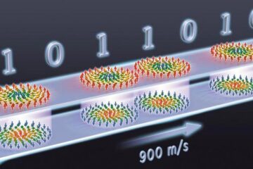
… a step towards the computing of the future. An international research team led by scientists from the CNRS1 has discovered that the magnetic nanobubbles2 known as skyrmions can be…


