Novel High-Resolution Methods in Fluorescence Microscopy
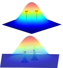
<br>The fluorescent signals from two nearby objects are superimposed by diffraction and imaged as a single feature. The ability to image individual probes separately means that their positions can be determined much more accurately to reconstruct the whole structure.<br>
Physical limits in high-resolution light microscopy can be overcome with the aid of chemical reactions. Scientists at Heidelberg University’s Institute of Physical Chemistry and members of the Cluster of Excellence “CellNetworks” have devised a new method in which light-dependent processes are replaced by chemical reactions to mark cellular structures for high-resolution optical microscopy. This method opens up new application vistas for fluorescence microscopy. The findings have been published online in the journal “Angewandte Chemie International Edition”.
Fluorescence microscopy is a widely used method for studying cellular structures. However, the so-called diffraction limit prevents detailed examination of these structures. Objects separated by less than 0.3 μm from one another cannot be resolved. Recently, different methods have been reported to overcome this limit, among them stochastic optical reconstruction microscopy (STORM). Here the cell structures are labelled with fluorescent dyes that respond with fluorescence emission upon stimulation by light of a certain wavelength. High resolution of approx. 0.02 μm can be achieved by switching off the majority of the dyes and leaving only a small number of them on. In this way, the light emitted by neighbouring dyes is no longer superimposed. This dye-switching is also controlled by light. The dyes left on can now be individually resolved and their positions can be determined by mathematical analysis to a very high accuracy of approx. 0.003 μm. Multiple repetition of this procedure supplies precise information about the location of all the dyes, thus permitting high-resolution reconstruction of the cell structures under investigation.
This method puts several demands on the microscope and the light sources used. Switching the respective dyes requires either different laser lines (excitation wavelength) or high light intensities or both. This can lead to problems in the investigation of living cells. Accordingly, the team headed by Heidelberg chemist Dr. Dirk-Peter Herten has replaced the switching of dyes via laser light with a light-independent process. To this end, the scientists adjusted a chemical probe for the detection of copper ions in such a way that the probe and its fluorescent properties can be used to mark cellular structures. If copper(II) binds to the probe, its fluorescence is suppressed. This binding of the copper(II) ion is reversible, thus restoring the fluorescence of the probe. The microscopic investigation of cell structures is therefore controlled by a reversible chemical reaction.
The scientists have called this method CHIRON – chemically improved resolution for optical nanoscopy. According to Dr. Herten, it means that microscopy methods like STORM can be simplified to such an extent that additional laser lines and high light intensities become superfluous. All that is required is for the probe to be located in a cellular environment to which minimal amounts of copper sulphate can be added, e.g. fixed cells. “This opens up new applications for high-resolution microscopy that were formerly impracticable due to technical restrictions,” says Dr. Herten. “Our probes can be used on many microscopes.”
For more information, go to
http://www.bioquant.uni-heidelberg.de/research/groups/single_molecule_spectroscopy.html.
Note for news desks:
Digital photo material is available from the Press Office.
Original publication
M. Schwering, A. Kiel, A. Kurz, K. Lymperopoulos, A. Sprödefeld, R. Krämer, D.-P. Herten: Far-Field Nanoscopy with Reversible Chemical Reactions / Hochauflösende Mikroskopie mit reversiblen chemischen Reaktionen. Angewandte Chemie International Edition, 15 February 2011, doi: 10.1002/anie.201006013
Contact:
Dr. Dirk-Peter Herten
Cluster of Excellence “CellNetworks”
Institute of Physical Chemistry
phone: +49 6221 5451220
dirk-peter.herten@urz.uni-hd.de
Communications and Marketing
Press Office, phone: +49 6221 542311
presse@rektorat.uni-heidelberg.de
Media Contact
All latest news from the category: Physics and Astronomy
This area deals with the fundamental laws and building blocks of nature and how they interact, the properties and the behavior of matter, and research into space and time and their structures.
innovations-report provides in-depth reports and articles on subjects such as astrophysics, laser technologies, nuclear, quantum, particle and solid-state physics, nanotechnologies, planetary research and findings (Mars, Venus) and developments related to the Hubble Telescope.


Newest articles

Silicon Carbide Innovation Alliance to drive industrial-scale semiconductor work
Known for its ability to withstand extreme environments and high voltages, silicon carbide (SiC) is a semiconducting material made up of silicon and carbon atoms arranged into crystals that is…

New SPECT/CT technique shows impressive biomarker identification
…offers increased access for prostate cancer patients. A novel SPECT/CT acquisition method can accurately detect radiopharmaceutical biodistribution in a convenient manner for prostate cancer patients, opening the door for more…

How 3D printers can give robots a soft touch
Soft skin coverings and touch sensors have emerged as a promising feature for robots that are both safer and more intuitive for human interaction, but they are expensive and difficult…

