3D bioprinting technique controls cell orientation

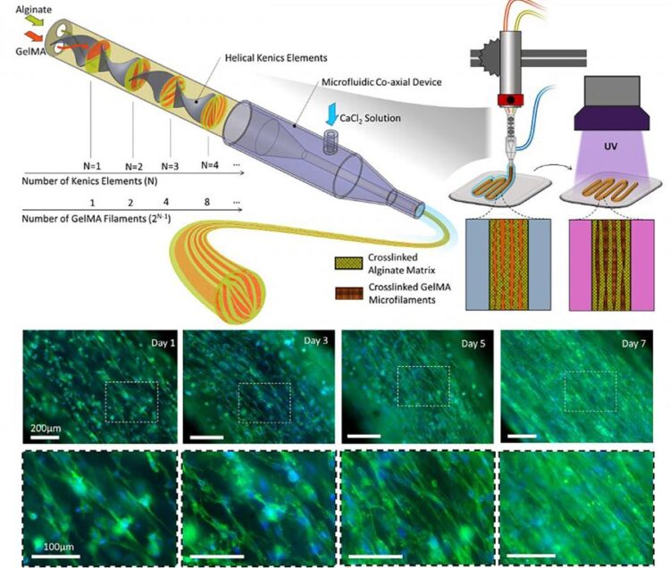
Biofabrication of multicompartmental hydrogel fibers for formation of multiscale biomimetic constructs.
Credit: Mohamadmahdi Samandari, Fatemeh Alipanah, Keivan Majidzadeh-A, Mario M. Alvarez, Grissel Trujillo-de Santiago, and Ali Tamayol
Common bioprinting methods fail to direct cell orientation at the individual cell level, but a technique can with implications for engineering skeletal muscles, tendons, and ligaments.
3D bioprinting can create engineered scaffolds that mimic natural tissue. Controlling the cellular organization within those engineered scaffolds for regenerative applications is a complex and challenging process.
Cell tissues tend to be highly ordered in terms of spatial distribution and alignment, so bioengineered cellular scaffolds for tissue engineering applications must closely resemble this orientation to be able to perform like natural tissue.
In Applied Physics Reviews, from AIP Publishing, an international research team describes its approach for directing cell orientation within deposited hydrogel fibers via a method called multicompartmental bioprinting.
The team uses static mixing to fabricate striated hydrogel fibers formed from packed microfilaments of different hydrogels. In this structure, some compartments provide a favorable environment for cell proliferation, while others act as morphological cues directing cell alignment. The millimeter-scale printed fiber with the microscale topology can rapidly organize the cells toward faster maturation of the engineered tissue.
“This strategy works on two principles,” said Ali Tamayol, coauthor and an associate professor in biological engineering at UConn Health. “The formation of topographies is based on the design of fluid within nozzles and controlled mixing of two separate precursors. After crosslinking, the interfaces of the two materials serve as 3D surfaces to provide topographical cues to cells encapsulated within the cell permissive compartment.”
Extrusion-based bioprinting is the most widely used bioprinting method. In extrusion-based bioprinting, the printed fibers are typically several hundreds of micrometers in size with randomly oriented cells, so a technique providing topographical cues to the cells within these fibers to direct their organization is highly desirable.
Conventional extrusion bioprinting also suffers from high shear stress applied to the cells during the extrusion of fine filaments. But the fine scale features of the proposed technique are passive and do not compromise other parameters of the printing process.
To direct cellular organization, according to the team, extrusion-based 3D-bioprinted scaffolds should be made from very fine filaments.
“It makes the process challenging and limits its biocompatibility and the number of materials that can be used, but with this strategy larger filaments can still direct cellular organization,” said Tamayol.
This bioprinting technique “enables production of tissue structures’ morphological features — with a resolution up to sizes comparable to the cells’ dimension — to control cellular behavior and form biomimetic structures,” Tamayol said. “And it shows great potential for engineering fibrillar tissues such as skeletal muscles, tendons, and ligaments.”
###
The article, “Controlling cellular organization in bioprinting through designed 3D microcompartmentalization,” is authored by Mohamadmahdi Samandari, Fatemeh Alipanah, Keivan Majidzadeh-A, Mario M. Alvarez, Grissel Trujillo-de Santiago, and Ali Tamayol. The article appears in Applied Physics Reviews (DOI: 10.1063/5.0040732) and can be accessed at https:/
All latest news from the category: Physics and Astronomy
This area deals with the fundamental laws and building blocks of nature and how they interact, the properties and the behavior of matter, and research into space and time and their structures.
innovations-report provides in-depth reports and articles on subjects such as astrophysics, laser technologies, nuclear, quantum, particle and solid-state physics, nanotechnologies, planetary research and findings (Mars, Venus) and developments related to the Hubble Telescope.
Newest articles

High-energy-density aqueous battery based on halogen multi-electron transfer
Traditional non-aqueous lithium-ion batteries have a high energy density, but their safety is compromised due to the flammable organic electrolytes they utilize. Aqueous batteries use water as the solvent for…

First-ever combined heart pump and pig kidney transplant
…gives new hope to patient with terminal illness. Surgeons at NYU Langone Health performed the first-ever combined mechanical heart pump and gene-edited pig kidney transplant surgery in a 54-year-old woman…

Biophysics: Testing how well biomarkers work
LMU researchers have developed a method to determine how reliably target proteins can be labeled using super-resolution fluorescence microscopy. Modern microscopy techniques make it possible to examine the inner workings…


