Ultrasound color Doppler twinkling in commercial breast biopsy markers
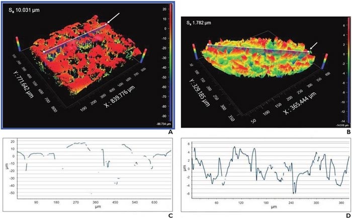
Analysis performed after removing general form and waviness from imaging data. Surface relief maps of (A) Cork and (B) MRI (Flex) markers. X-Y plots of corresponding slice profile for (C) Cork and (D) MRI Flex markers. Arrow in surface relief maps shows slice location for X-Y plots. Discontinuities in curves indicate regions where light from instrument was not adequately reflected to measure surface.
Credit: ARRS/AJR
Ultrasound color Doppler twinkling may aid the detection of certain biopsy markers in metastatic axillary nodes that resume normal morphology after neoadjuvant systemic therapy.
According to ARRS’ American Journal of Roentgenology (AJR), ultrasound color Doppler twinkling may aid the detection of certain biopsy markers in metastatic axillary nodes that resume normal morphology after neoadjuvant systemic therapy.
Noting that certain breast biopsy markers exhibited actionable twinkling (i.e., sufficient confidence to rely solely on twinkling for target localization) in cadaveric breast, “twinkling was observed with greater confidence for C1-6 and 9L than ML6-15 transducer,” wrote first author Christine U. Lee, MD, PhD, from the department of radiology at Mayo Clinic in Rochester, MN. “Actionable twinkling was associated with higher marker surface roughness.”
Dr. Lee and colleagues evaluated 35 commercial breast biopsy markers for twinkling artifact in various experimental conditions, including scanning medium (solid gel phantom, ultrasound coupling gel, cadaveric breast), transducer (ML6-15, 9L, C1-6), and embedding material (present vs. absent). Markers were then assigned twinkling scores from 0 (confident in no twinkling) to 4 (confident in exuberant twinkling); score ≥3 represented actionable twinkling.
Ultimately, three breast biopsy markers—Cork, Q, and MRI (Flex)—exhibited actionable ultrasound twinkling for ≥2 transducers in cadaveric breast. Additionally, surface roughness was significantly higher for markers with than without actionable twinkling for the C1-6 (median values: 0.97 vs 0.35, p=.02) and 9L (1.75 vs. 0.36; p=.002) transducers.
“The use of twinkling artifact to help detect biopsy markers by ultrasound could impact breast radiologists in performing preoperative localization procedures after NST,” the authors of this AJR article concluded, thus impacting breast surgeons in performing intraoperative localization.
An electronic supplement to this AJR article is available here.
North America’s first radiological society, the American Roentgen Ray Society (ARRS) remains dedicated to the advancement of medicine through the profession of medical imaging and its allied sciences. An international forum for progress in radiology since the discovery of the x-ray, ARRS maintains its mission of improving health through a community committed to advancing knowledge and skills with the world’s longest continuously published radiology journal—American Journal of Roentgenology—the ARRS Annual Meeting, InPractice magazine, topical symposia, myriad multimedia educational materials, as well as awarding scholarships via The Roentgen Fund®.
Journal: American Journal of Roentgenology
DOI: 10.2214/AJR.22.28107
Method of Research: Imaging analysis
Subject of Research: People
Article Title: Factors associated with ultrasound color Doppler twinkling by breast biopsy markers: In vitro and ex vivo evaluation of 35 commercially available markers
Article Publication Date: 31-Aug-2022
Media Contact
Logan Young
American Roentgen Ray Society
lyoung@arrs.org
Office: 703-858-4332
All latest news from the category: Medical Engineering
The development of medical equipment, products and technical procedures is characterized by high research and development costs in a variety of fields related to the study of human medicine.
innovations-report provides informative and stimulating reports and articles on topics ranging from imaging processes, cell and tissue techniques, optical techniques, implants, orthopedic aids, clinical and medical office equipment, dialysis systems and x-ray/radiation monitoring devices to endoscopy, ultrasound, surgical techniques, and dental materials.


Newest articles

Superradiant atoms could push the boundaries of how precisely time can be measured
Superradiant atoms can help us measure time more precisely than ever. In a new study, researchers from the University of Copenhagen present a new method for measuring the time interval,…

Ion thermoelectric conversion devices for near room temperature
The electrode sheet of the thermoelectric device consists of ionic hydrogel, which is sandwiched between the electrodes to form, and the Prussian blue on the electrode undergoes a redox reaction…

Zap Energy achieves 37-million-degree temperatures in a compact device
New publication reports record electron temperatures for a small-scale, sheared-flow-stabilized Z-pinch fusion device. In the nine decades since humans first produced fusion reactions, only a few fusion technologies have demonstrated…

